
Prurigo Nodularis (Hyde’s Prurigo)

What Are Chronic Oral Mucosal Lesions?
Chronic lesions of the oral mucosa are disorders of the mouth that are characterized by soreness and sores of the soft mucosal lining of the mouth.
Chronic lesions of the oral mucosa are disorders of the mouth that are characterized by soreness and sores of the soft mucosal lining of the mouth.
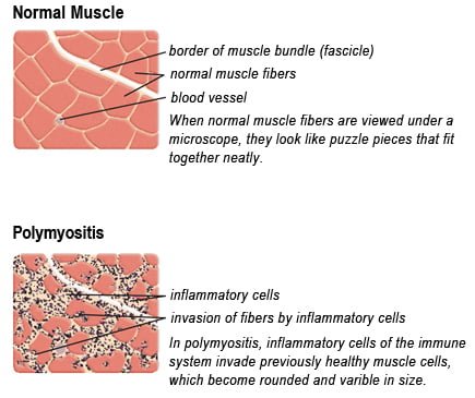
P olymyositis is a body-wide connective tissue disease resulting from autoimmune attack of skeletal muscles that is characterized by inflammatory and degeneratory changes. The course is unpredictable being marked by spontaneous flare-ups and remissions.
Polymyositis can begin slowly or abruptly according to the factor that is triggering the onset such as infection, medications like phenytoin, and autoimmune disease.
Progressive muscle weakness starts in the proximal skeletal muscles (muscles closest to the trunk of the body).
Skeletal muscles, also called voluntary, are muscles that move the body as we want, such as walking and lifting objects, as opposed to those we cannot voluntarily control, such as the muscles of digestion.
Q: What are the degeneratory changes in skeletal muscles?
A: In polymyositis, degeneratory changes in skeletal muscles means that muscles are being destroyed (called necrosis), resulting in fibrosis, or scarring. When scar tissue takes the place of lost muscle tissue, it cannot act like muscle to contract and relax. Muscle destruction is what causes muscle pain and weakness.
After the clinical work-up of exams and blood studies to determine muscle damage, the diagnosis of polymyositis is confirmed by muscle biopsy. See image at above left. The black dots are inflammatory cells. Edema (fluid) between cells caused by inflammation pushes muscle fibers apart.
There is no cure for polymyositis, but the symptoms can be treated. Options include medication, physical therapy, exercise, heat therapy (including microwave and ultrasound), orthotics and assistive devices, and rest. The standard treatment for polymyositis is a corticosteroid drug, given either in pill form or intravenously. Immunosuppressant drugs, such as azathioprine and methotrexate, may reduce inflammation in people who do not respond well to prednisone.
Periodic treatment using intravenous immunoglobulin can also improve recovery. Other immunosuppressive agents used to treat the inflammation associated with polymyositis include cyclosporine A, cyclophosphamide, and tacrolimus. Physical therapy is usually recommended to prevent muscle atrophy and to regain muscle strength and range of motion.1
Diagnosis is based on elevated muscles enzymes, increased urinary creatine level, and electromyograph abnormalities.
Polymyositis can affect people at any age. It is most common in adults between ages 50 and 70, and in children ages 5 to 15. It affects women twice as often as men and is more common in African Americans than Caucasians.2 The major causes of death from polymyositis are cancer and lung disease, including pneumonia. The 5-year mortality rate can be as high as 1 in 5 patients.2
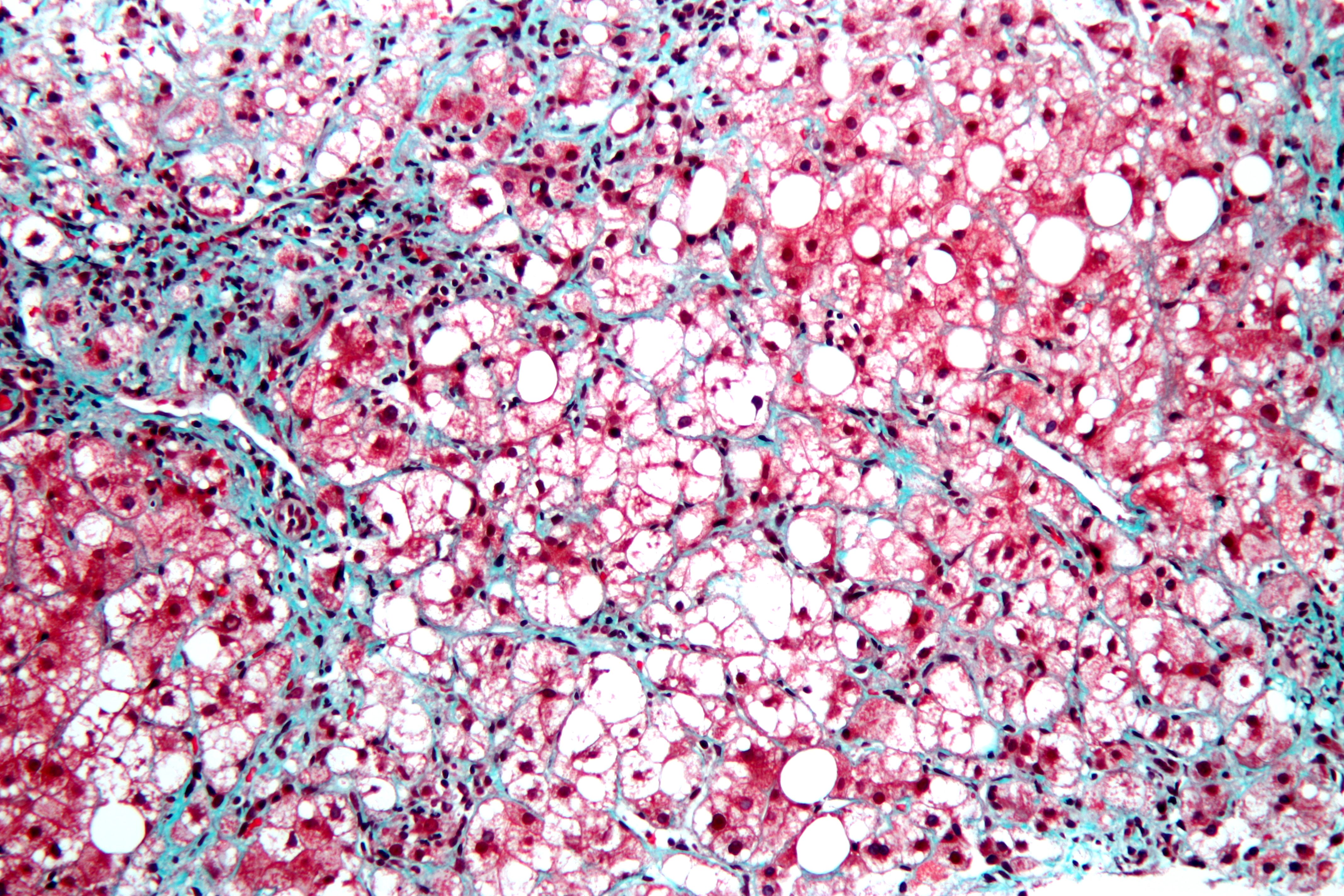
N on-alcoholic fatty liver is a non-inflammatory liver disorder characterized by degenerative changes in the liver caused by excessive accumulation of lipid (fat) in hepatocytes (liver cells) that is called free fatty acid-generated lipotoxicity.
Non-alcoholic fatty liver shows an increase in liver enzymes called transaminases.
Q: What are the enzymes that increase?
A: The transaminases that increase are ALT and AST. ALT is the abbreviation for alanine aminotransferase enzyme and AST is the abbreviation for aspartate aminotransferase enzyme. They are commonly measured in blood tests to determine liver function and when elevated indicate inflammation.

P rurigo nodularis is a chronic dermatitis characterized by hard, dry, deep seated, intensely itchy papules (small bumps like pimples) and/or nodules (large bumps) that erupt most commonly on the arms, legs, and back.
Papules and nodules vary in number and may become infected after picking or scratching.
Q: Does the itching go away?
A: New nodules develop from time to time, and existing nodules may remain itchy indefinitely, although some may regress spontaneously and leave scars. In most cases, the disease runs a very protracted course with exacerbations and remissions.1
Prurigo nodularis is an unusual disorder of unknown etiology, which is notoriously resistant to therapy. A variety of systemic conditions have been reported to be associated with prurigo nodularis. However, the mechanism by which these disorders may trigger prurigo nodularis is unknown.2
It has been shown to be associated with malnutriton and infection such as tonsillitis, which resolved after removal of tonsils.3
 What Is Hypokalemic Rhabdomyolysis?
H ypokalemic rhabdomyolysis is an acute and sometimes fatal disease due to its rapid progression of muscle destruction when untreated.
It is characterized by the accumulation of by-products of skeletal muscle destruction in the renal (kidney) tubules and producing acute kidney failure caused by rapid potassium loss.
This condition puts you in bed because the legs muscles cannot support the body and arms are too weak to move.

P ancreatic insufficiency is a disorder characterized by insufficient exocrine production of pancreatic enzymes for normal digestion of fats, proteins, and carbohydrates that results in maldigestion of these foodstuffs.
Pancreatic insufficiency also causes malabsorption of the fat-soluble vitamins: vitamin A, vitamin D, vitamin E, and vitamin K.
Q: What are the exocrine enzymes produced by the pancreas?
A: Exocrine enzymes produced by the pancreas include amylase for the digestion of carbohydrates, lipase for the digestion of fats, and protease for the digestion of proteins.
While lipases and amylase are secreted in the active form, proteases are secreted as pro-enzymes (need to be activated). Trypsinogen is converted to its active form trypsin in the duodenum by enterokinase, a protease secreted by the enterocytes (surface cells that line the duodenum), and trypsin in turn activates the other pancreatic proteases.1
Pancreatic enzymes are released by the pancreas into the internal pancreatic duct that empties into the common bile duct from which they are pumped through the Sphincter of Odi directly into the duodenum as needed. Tthe Sphincter of Odi is a strong circular muscle that controls the entrance of pancreatic enzymes into the duodenum.
After a meal, enzyme secretion into the duodenum increases quickly reaching peak output within the first 20 to 60 minutes, then decreasing to a stable level before reaching an interdigestive level at the end of the digestive period, that is, about 4 hours after meal intake.2
Medical treatment is with medication that contains enzymes to break down carbohydrates, fats, and protein in food at mealtime. This treatment is safe, effective, and has few side effects.
S teatorrhea is a condition of abnormal stool composition and consistency that is due to high fat content because fat from food is not digested and absorbed into the body but rather passes out with stool.1
Q: How does fatty stool look?
A: The stool in steatorrhea appears pale, bulky and may float in the toilet. If stool is loose, it varies from a pudding consistency to whipped cream. If it is solid, it can vary from thin sqiggles to big turds that smear the sides of the toilet bowl. All types have a foul smell.
Fat of any kind must be digested, or broken down, by enzymes into fatty acids and glycerol and then these parts only are absorbed through the small intestinal lining into the body. The failure to digest or absorb fats deprives the body of these foodstuffs needed for many functions such as blood clotting and vital cell parts such as eye and brain structure.
Fat malabsorption produces malabsorption of vitamins A, D, E, and K because these vitamins can only be absorbed along with fatty acids.

R ounded, curved, dark and dry nails are a well known feature of vitamin B12 deficiency.
Q: How does vitamin B12 deficiency cause the nails to grow this way?
A: Vitamin B12 deficiency affects the nail plate. The nail plate is the hard keratin cover of the finger tip and toe tip, simply called “the nail.” The nail plate is produced by the living nail matrix at its base.

T ype 1 diabetes mellitus (T1DM), also termed type 1A, is an inherited autoimmune disorder in which anti-islet autoantibodies destroy the islet cells of the pancreas that secrete insulin hormone. Type 1 diabetes mellitus was formerly called juvenile diabetes because it usually afflicts persons under the age of 25 years.
Loss of insulin production results in failure to metabolize glucose. Glucose is a simple sugar that is a required source of energy for the body, especially the brain and muscles.
Type 1 diabetes mellitus is characterized by sustained fasting blood glucose levels above 126 mg/dL (hyperglycemia) with subsequent loss of glucose from the body by removal through the urine (glucosuria) as the body attempts to lower blood glucose, and cell starvation that follows.
That is, while glucose accumulates in blood, the body cannot access it. Without insulin treatment, this disorder quickly produces coma and ultimately results in death. In fact, it is 5th leading cause of death in the United States.
Q: How does insulin work?
A: Insulin moves glucose from the bloodstream into body cells where it is used or reformulated for high energy storage. For example, muscles can use glucose for immediate work or store it in the form of glygogen for later work, depending on need. Healthy insulin production keeps an 8 hour fasting blood glucose level to less than 100 mg/dL. Upon eating carbohydrate food, glucose is digested and absorbed from the small intestine into the bloodstream which then raises blood glucose levels. The elevated level is controlled by prompt action of insulin to lower it to below 140 mg/dL within 2 hours of eating.
Insulin does not work alone. The islets of Langerhans manage glucose in the body. The islets are specialized formations located on the outer surface of the pancreas. The islets are composed of two different types of cells known as alpha and beta cells. These cells make the competing hormones that keep blood glucose within a healthy range.
Alpha cells secrete glucagon to raise blood glucose levels by triggering the body to release stored energy in the form of glycogen. In the opposite, beta cells secrete insulin to lower blood glucose by opening body cells so that glucose in blood can enter. Without insulin, glucose cannot enter cells but remains in the bloodstream where it accumulates.
Insulin is also needed to move magnesium into cells from the bloodstream. On the other side, magnesium is needed to produce insulin. Insulin has other functions such as building muscle and helping regulate cholesterol which directly impacts the sex hormones, estrogen, progesterone, and testosterone.
Onset of symptoms usually occurs over a period of days or weeks, although beta cell destruction can begin years earlier. The SEARCH for Diabetes in Youth multicenter study, funded by the Centers for Disease Control and Prevention (CDC) and the National Institutes of Health (NIH), has determined that based on data from 2002 to 2003, a total of 15,000 youth in the United States were newly diagnosed with type 1 diabetes each year. Non-Hispanic white youth had the highest rate of new cases of type 1 diabetes according to NIH.
Type 1A diabetes mellitus has become one of the most intensively studied autoimmune disorders. It is now possible to predict its development, beginning with HLA-encoded genetic susceptibility, followed by the development of a series of anti-islet autoantibodies.1

C oronary artery disease (CAD), also called ischemic heart disease, is a gradual narrowing of medium and large arteries of the heart by fatty buildups, called atherosclerotic plaques.
It is characterized by slowly developing interference with blood flow to heart tissue itself, resulting in oppressive chest pain called angina and, ultimately, thrombosis (clot) causing heart attack.
The heart is a muscular organ that is working all the time, so it needs a constant supply of oxygen. Oxygen is brought to the working heart tissue by the coronary arteries with each beat of the heart. When heart muscle has to work harder, it needs more oxygen delivered to itself. Lack of oxygen causes pain.
In fact, failure of diseased coronary arteries to deliver adequate oxygen to heart tissue is the most common cause of angina pectoris – substernal pain (under breastbone) or pressure brought on by exertion and relieved by rest.
Thrombosis, or clot formation, occurs when blood cells within a narrowed artery can no longer get through. Trapped, blood cells pile up and block the artery thus triggering a cascade of events called heart attack. Coronary arteries that are narrowed by atherosclerotic plaques can rupture causing injury to the coronary blood vessel resulting in blood clotting which blocks the flow of blood to the heart muscle. Blood clots may form, partially dissolve, and later form again and angina can occur each time a clot blocks blood flow in an artery.1
Q: How does coronary artery disease develop?
A: Coronary artery disease slowly develops from this combination of events:
Dysfunction of epithelial cells that line the inside of arteries cause the vessels to stiffen, and subsequently
Accumulation of lipid (fat) in smooth muscle cells beneath the inside lining of arteries and in foam cells cause buildup of fatty deposits on the inside walls progressing to fibrous plaque formation.
Oxidized low-density lipoprotein (oxLDL), so-called bad cholesterol, and oxysterols play important roles in the development of atherosclerosis. OxLDL triggers the immune system to produce autoantibodies against oxLDL that are detectable in serum. These antibodies are called anti-oxLDL. Anti-oxLDL antibody and oxysterol concentrations are associated with coronary artery stenosis. Oxidative stress may be greatly increased in unstable angina.2 and Chronic inflammation in the general population is a major risk factor for ischemic heart disease.
The pathophysiology of atherosclerosis is, clearly, different in women when compared to the men. The women have a higher risk of blood coagulability making them at high risk for the blood clot formation. In a large number of women endothelial dysfunction, small vessel size and diffuse atherosclerosis have been identified as causes of ischemia without evidence of blockade in the coronary arteries.3
Also, atherosclerotic plaque in women is less fibrotic and contains more lipid filled foam cells, implying greater potential for reversibility but also potentially greater vulnerability for plaque rupture and thrombosis.4
Who is Affected in the General Population?
Coronary artery disease remains the leading cause of death in developed countries despite significant progress in primary prevention and treatment strategies.
It is the leading cause of death in women, as well as an important cause of disability.
Older patients are at particularly high risk of poor outcomes following acute coronary syndrome.5
Ischemic heart disease is the leading cause of death in the United States, making cardiovascular risk assessments and potential interventions or treatments imperative for patients with celiac disease.6
Sources: