Skip to content

Systemic Lupus Erythematosus 

bruiseWhat Is Hypoprothrombinemia?

[dropcap]H ypoprothrombinemia is a deficiency of prothrombin (clotting factor II) in the blood that is characterized by impaired hemostasis in response to trauma or a laceration.

Q: What is hemostasis and how is it altered by a deficiency of prothrombin?

A: Hemostasis encompasses the tightly regulated processes of blood clotting, platelet activation, and blood vessel repair.1

Prothrombin is a protein clotting factor present in blood that is involved in the first part of hemostasis, which is blood clotting or coagulation. Vitamin K is required for prothrombin production.

When a laceration or wound is sustained, prothrombin is converted to the enzyme thrombin. Thrombin in turn acts on fibrinogen to convert it to fibrin which then forms the framework of a clot to stop bleeding. Deficiency of prothrombin prevents this series of events and bleeding is not properly stopped.

After the clotting process of hemostasis would come the second part, platelet activation. Eventually, coagulation and platelet activation are switched off by blood-borne inhibitors.

What Is Hypoprothrombinemia In Celiac Disease and/or Gluten Sensitivity?

Sources:
  1. Versteeg HH, Heemskerk JWM, Levi M,  Reitsma PH. New Fundamentals in Hemostasis. Physiological Reviews Published 1 January 2013Vol. 93no. 327-358DOI: 10.1152/physrev.00016.2011 []

Sarcoidosis

What Is Vitamin B12 Deficiency Anemia? [dropcap]V itamin B12 deficiency anemia is a megaloblastic anemia that is characterized by defective DNA synthesis of red blood cells due to a lack of vitamin B12. Vitamin B12 is… 

Multiple Sclerosis

canstockphoto17997339What Is Gluten Sensitive Enteropathy?

Gluten sensitive enteropathy is active celiac disease characterized by inflammation of the small intestinal mucosa that results from an inherited immunologic intolerance to ingested gluten.

Q: What does the inflammation do to the mucosa in the small intestine?

A: Inflammation is a cell level immune response to gluten that has these effects on the mucosa:

  • Damages the barely visible villi (multitudinous finger-like structures) by causing atrophy or loss.
  • Likely affects the structural support and microcirculation of the villus, leading to collapse of the villus.
  • Elongates the crypts between villi. The thickening of the crypt is not so much a response to loss of surface enterocytes but represents inflammation of the mucosa.1
  • Increases round cells in the lamina propria and surface epithelial cells leaving few, irregular microvilli (brush border) on the surface of villi.
  • Damage is most intense in the duodenum and decreases toward the large intestine.
  • The extent of the damage to the intestine determines the malabsorptive consequences of the disease. Both gastric and small intestinal permeability are disrupted in patients with celiac disease.2
  • Relationship between active celiac disease and intestinal permeability: There is a clear association between degree of mucosal damage and the intestinal-permeability ratio, and a normal ratio generally implies near-normal small intestinal structure. A raised intestinal permeability of the mucosal lining (leaky gut) could predispose to a high absorption of gluten and exacerbate an existing lesion and hence convert a latent to an overt enteropathy.3
  • Relationship between active celiac disease and tight junction proteins: A study of intestinal permeability showed that the expression of all junction proteins of the small intestinal lining (occludin, claudin 3, zonula occludens 1, and E-cadherin) was already decreased in early stage celiac disease when compared with non-celiac controls, showing leaky gut and confirming the above earlier study by Johnston et al. Junction protein expression correlated positively with mucosal villus structure and negatively with the number of intraepithelial lymphocytes (IELs), the intensity of small-intestinal autoantibody deposits, and serum autoantibodies. The expression of claudin 3 showed a negative correlation with diarrheal score.4
  • Relationship between active celiac disease and inflammation. In celiac disease there is an over production of inflammatory interleukin-15 (IL-15) which inhibits the correct removal of damaged intraepithelial lymphocytes caused by the reaction to gluten. Serum levels of IL-15 are directly correlated with the seriousness of tissue damage.5
  • Relationship between active celiac disease and gut microbiota. Results of a study investigating intestinal microbiota (normal bacterial residents) in patients with celiac disease suggest that with lower levels of the genus bifidobacteria, celiac patients have an imbalance in the intestinal microbiota even while on a gluten-free diet. This fact could favor the pathological process of the disorder. The concentration of bifidobacteria per gram of feces was significantly higher in healthy subjects (2.5 ± 1.5 x10⁷ CFU/g) when compared to celiac patients (1.5 ± 0.63 x10⁸ CFU/g).6

  • Relationship between active celiac disease and endoscopy technique. The most severe degree of villous atrophy was detected when distal duodenal biopsy specimens were taken in addition to a duodenal bulb biopsy specimen from either the 9- or 12-o’clock position (96.4% sensitivity; 95% CI, 79.7%-100%). The difference between the 12-o’clock position biopsy and the 3-o’clock position biopsy in detecting the most severe villous atrophy was 92% (24/26 patients) versus 65% (17/26 patients).7
  • Relationship between active celiac disease and diet adherence. Patients with consistent gluten free diet adherence experience symptomatic responses to dietary gluten (SRDG) faster and more severe in comparison to their prior gluten exposure possibly demonstrating an adept immunological response. Anxiety and depression also enhance the speed of symptom onset and co-existing visceral hypersensitivity is a risk factor for severe reactions to dietary gluten.8
  • Relationship between active celiac disease and atrial fibrillation: Patients with celiac disease, verified by intestinal biopsy, are at increased risk of atrial fibrillation. This observation is consistent with previous findings that elevation of inflammatory markers predicts atrial fibrillation.9

How Prevalent Is Gluten Sensitive Enteropathy?

Sources:
  1. Murray JA, the widening spectrum of celiac disease. American Journal of Clinical Nutrition. Mar 1999; 69(3):354-365. []
  2. Murray JA, the widening spectrum of celiac disease. American Journal of Clinical Nutrition. Mar 1999; 69(3):354-365. []
  3. Johnston SD, Smye M, Watson RGP. Intestinal permeability and morphometric recovery in coeliac disease. Lancet. Jul 28, 2001;358(9278):259, 2p. []
  4. Rauhavirta T, Lindfors K, Koskinen O, Laurila K, Kurppa K, Saavalainen P, Mäki M, Collin P, Kaukinen K. Impaired epithelial integrity in the duodenal mucosa in early stages of celiac disease. Transl Res. 2014 Sep;164(3):223-31. doi: 10.1016/j.trsl.2014.02.006 []
  5. Stazi AV, Trinti B. Selenium status and over-expression of interleukin-15 in celiac disease and autoimmune thyroid diseases. Ann Ist Super Sanita. 2010;46(4):389-99.DOI: 10.4415/ANN_10_04_06. []
  6. Golfetto L, de Senna FD, Hermes J, Beserra BT, França Fda S, Martinello F. Lower bifidobacteria counts in adult patients with celiac disease on a gluten-free diet. Arq Gastroenterol. 2014 Apr-Jun;51(2):139-43. []
  7. Kurien M, Evans KE, Hopper AD, Hale MF, Cross SS, Sanders DS. Duodenal bulb biopsies for diagnosing adult celiac disease: is there an optimal biopsy site? Gastrointest Endosc. 2012 Jun;75(6):1190-6. doi: 10.1016/j.gie.2012.02.025. []
  8. Barratt SM, Leeds JS, Sanders DS. Factors influencing the type, timing and severity of symptomatic responses to dietary gluten in patients with biopsy-proven coeliac disease. J Gastrointestin Liver Dis. 2013 Dec;22(4):391-6. []
  9. Emilsson L, Smith JG, West J, Melander O, Ludvigsson JF. Increased risk of atrial fibrillation in patients with coeliac disease: a nationwide cohort study. Eur Heart J. 2011 Oct;32(19):2430-7. doi: 10.1093/eurheartj/ehr167. []

Arthritis, Juvenile Idiopathic

Lymphadenopathy.
Lymphadenopathy affecting a node in the neck.

What Is Lymphadenopathy?

[dropcap]L ymphadenopathy is an alteration of lymph nodes that is characterized by enlargement of lymph nodes greater than 1.5 cm caused by proliferation (increased production) of lymphocytes within the node.

Q: What are lymph nodes?

A: Lymph nodes are part of the lymphatic system, acting to protect body fluids by filtering out and destroying bacteria and other harmful substances from lymph that is continually carried to them by lymph vessels.

Cleaned lymph is carried away from the nodes by lymph vessels to the bloodsteam where it helps forms blood plasma. Lymph nodes produce various blood cells needed to fight infection which includes lymphocytes.

Lymphocytes are small white blood cells that plays a major role in defending the body against disease. There are two types of lynphocytes: B cells, which make antibodies that attack bacteria and toxins, and T cells which attack body cells themselves when they have been taken over by viruses or become cancerous.

Lymph nodes that become enlarged doing battle with an infection or as a result of injury nearby usually resolve with treatment of the infection or injury. However, if the cause is cancer, the nodes would need to be treated as well as the cancer.

What Is Lymphadenopathy In Celiac Disease and/or Gluten Sensitivity?

Psoriasis

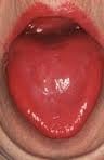
niacin deficiencyWhat Is A Fiery Red, Smooth, Burning Tongue?

[dropcap]A fiery red, smooth, burning tongue is an alteration in tongue tissue that is characteristic of advanced niacin deficiency.1

Q: How does niacin deficiency cause the tongue to be red and sore?

A: Niacin is an essential B vitamin that is required for a healthy tongue and by all body cells as well. Deficiency first shows in tissues with rapid cellular turnover, such as mucosal cells of the tongue.

When absorbed from the small intestines, niacin (the form in food) becomes the active form niacinamide. Niacinamide is converted by the body into co-enzymes which are present in all cells. These enzymes function in oxidation-reduction reactions essential for release of energy from carbohydrates, fats, and proteins and are needed as components for more than 200 enzymes involved in metabolism.

In addition to producing energy, niacinamide is essential for healthy skin and the mucosal lining of the digestive tract, normal functioning of the brain and nervous system, and production of steroid hormones from adrenal glands and hormones from sex glands.

What Is A Fiery Red, Smooth, Burning Tongue In Celiac Disease and/or Gluten Sensitivity?

Sources:
  1. Krause’s Food, Nutrition, & Diet Therapy. 10th Edition. Kathleen Mahan, Sylvia Escott-Stump. 2000. W.B. Saunders Company. []