This 21 year-old woman (right) appears as old as her 70 year-old grandmother (left). Courtesy Prof Dr Chua Chung Nen
What Is Acquired Cutis Laxa?
[dropcap]A cquired cutis laxa is an uncommon skin disorder characterized by abnormal reduction and degeneration of elastic fibers of the skin that can appear simply as thick, saggy skin with loose folds to severe involvement showing a premature aged appearance.
Q: What are elastic fibers of the skin?
A: Elastic fibers of the skin are connective tissue found in the dermis, which is the layer of skin under the epidermis, or surface layer. They hold the shape of skin and are important for wound healing in the development of scars.
What Is Cutis Laxa In Celiac Disease and/or Gluten Sensitivity?
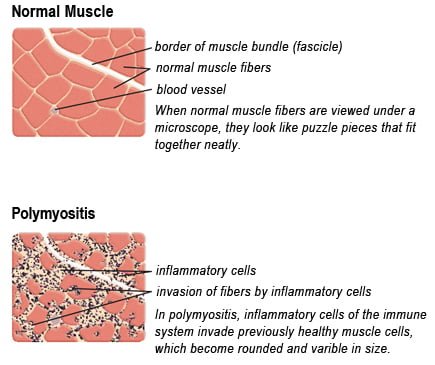
Drawing of Biopsy Showing Muscle Fibers Invaded by Immune Cells. Courtesy MDA.org
What Is Polymyositis?
[dropcap]P olymyositis is a body-wide connective tissue disease resulting from autoimmune attack of skeletal muscles that is characterized by inflammatory and degeneratory changes. The course is unpredictable being marked by spontaneous flare-ups and remissions.
Polymyositis can begin slowly or abruptly according to the factor that is triggering the onset such as infection, medications like phenytoin, and autoimmune disease.
Progressive muscle weakness starts in the proximal skeletal muscles (muscles closest to the trunk of the body).
Skeletal muscles, also called voluntary, are muscles that move the body as we want, such as walking and lifting objects, as opposed to those we cannot voluntarily control, such as the muscles of digestion.
Q: What are the degeneratory changes in skeletal muscles?
A: In polymyositis, degeneratory changes in skeletal muscles means that muscles are being destroyed (called necrosis), resulting in fibrosis, or scarring. When scar tissue takes the place of lost muscle tissue, it cannot act like muscle to contract and relax. Muscle destruction is what causes muscle pain and weakness.
After the clinical work-up of exams and blood studies to determine muscle damage, the diagnosis of polymyositis is confirmed by muscle biopsy. See image at above left. The black dots are inflammatory cells. Edema (fluid) between cells caused by inflammation pushes muscle fibers apart.
There is no cure for polymyositis, but the symptoms can be treated. Options include medication, physical therapy, exercise, heat therapy (including microwave and ultrasound), orthotics and assistive devices, and rest. The standard treatment for polymyositis is a corticosteroid drug, given either in pill form or intravenously. Immunosuppressant drugs, such as azathioprine and methotrexate, may reduce inflammation in people who do not respond well to prednisone.
Periodic treatment using intravenous immunoglobulin can also improve recovery. Other immunosuppressive agents used to treat the inflammation associated with polymyositis include cyclosporine A, cyclophosphamide, and tacrolimus. Physical therapy is usually recommended to prevent muscle atrophy and to regain muscle strength and range of motion.1
Diagnosis is based on elevated muscles enzymes, increased urinary creatine level, and electromyograph abnormalities.
Polymyositis can affect people at any age. It is most common in adults between ages 50 and 70, and in children ages 5 to 15. It affects women twice as often as men and is more common in African Americans than Caucasians.2 The major causes of death from polymyositis are cancer and lung disease, including pneumonia. The 5-year mortality rate can be as high as 1 in 5 patients.2
What Is Polymyositis In Celiac Disease and/or Gluten Sensitivity?
Sources:
National Institute of Neurological Disorders and Stroke [↩]
Nail Anatomy. A. Nail plate; B. lunula; C. root; D. sinus; E. matrix; F. nail bed; G. hyponychium; H. free margin. Courtesy Wikimedia.org
What Are Rounded Nails With Curved Ends?
[dropcap]R ounded, curved, dark and dry nails are a well known feature of vitamin B12 deficiency.
Q: How does vitamin B12 deficiency cause the nails to grow this way?
A: Vitamin B12 deficiency affects the nail plate. The nail plate is the hard keratin cover of the finger tip and toe tip, simply called “the nail.” The nail plate is produced by the living nail matrix at its base.
What Are Rounded Nails With Curved Ends In Celiac Disease and/or Gluten Sensitivity?
[dropcap]W hite spots in the nails is a feature of zinc deficiency and white bands signify protein deficiency characterized by abnormal appearance anywhere in one or more nails.
White spots and bands can appear separately or together in nails. There may be just one mark or many marks depending on the level of deficiency.
Q: What part of the nail is affected by zinc and/or protein deficiency?
A: Zinc and protein deficiencies affect the nail plate.
The nail plate is the hard keratin cover of the finger tip and toe tip, simply called “the nail.” The nail plate is produced by the living nail tissue at its base. Here is an illustration of the parts that make up a fingernail:
Nail Anatomy. .
A. Nail plate; B. lunula; C. root; D. sinus; E. matrix; F. nail bed; G. hyponychium; H. free margin. Courtesy Wikipedia.
What Are White Spots And White Bands In Nails In Celiac Disease and/or Gluten Sensitivity?
Cutaneous vasculitis is a vascular disease of small blood vessels serving the skin that is characterized by segmental (spotty) inflammation of affected vessel walls resulting in their necrosis (destruction). Scarring is seen in the affected skin that lies over the destroyed blood vessels.
Skin appearance varies from small flat, purplish spots to raised purplish areas over damaged vessels.
In most cases, cutaneous vasculitis results from deposits of toxic immune complexes in the affected vessel walls that is caused by autoimmune activity targeting small blood vessels.1
Cutaneous vasculitis can also result from infection, drug reactions, or malignancies.
Who is Affected in the general Population? Cutaneous vasculitis affects all ages.
The overall annual incidence of cutaneous vasculitis was 38.6 persons per million in 1998.2
What Is Cutaneous Vasculitis In Celiac Disease and/or Gluten Sensitivity?
Sources:
Abenavoli L, Proietti I, Leggio L, Ferrulli A, Vonghia L, Capizzi R, Rotoli M, Amerio PL, Gasbarrini G, Addolorato G. Cutaneous manifestations in celiac disease. World J Gastroenterol. 2006 Feb 14;12(6):843-52. [↩]
Watts RA, Jolliffe VA, Grattan CE, Elliott J, Lockwood M, Scott DG. Cutaneous vasculitis in a defined population–clinical and epidemiological associations. J Rheumatol. 1998 May;25(5):920-4. [↩]
Cancel your subscription?
We're sorry to see you go.
Please select a reason for cancellation.
Please provide additional details.
Cancellation request submitted
We've received your request. Your subscription remains active while we process it. We'll email you once it's confirmed.
Cancellation pending
Access Content
Affiliate Terms
By joining our Affiliate Program, you agree to the following terms:
1. Eligibility: You must be 18 years or older to participate. We reserve the right to refuse service to anyone for any reason at any time.
2. Promotional Guidelines: Affiliates must not engage in misleading advertising or spam. Promotions must accurately represent our brand and products.
3. Affiliate Links: You must use your unique tracking link to receive credit for referred sales.
4. Prohibited Use: You may not use our branding in a way that may confuse customers or misrepresent your relationship with us.
5. Termination: We reserve the right to terminate your account at any time for breach of these terms.
6. Changes to Agreement: We may update these terms at any time. Continued participation implies acceptance of any changes.
By continuing, you agree to the terms of this Affiliate Agreement.