Skip to content

Arachidonic Acid (AA) Deficiency
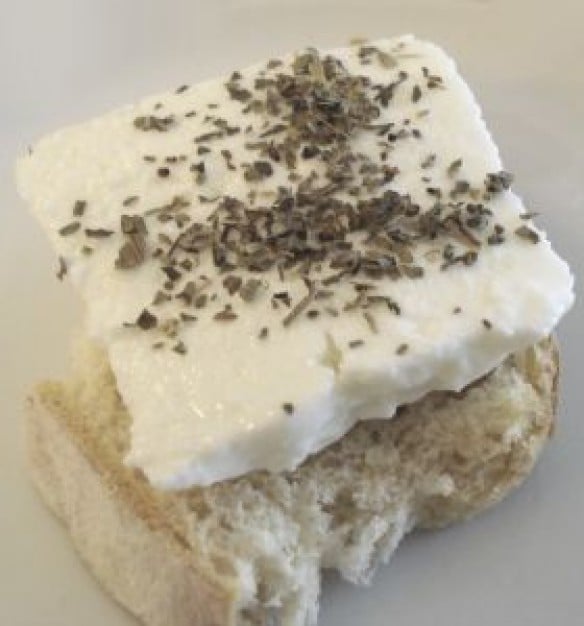
QueijoFresco.
QueijoFresco.

What Is Arachidonic Acid?

[dropcap]A[/dropcap]rachidonic acid is a major essential (must have/can’t make) omega-6 fatty acid.

Structurally, arachidonic acid is a key component of nerve membranes, together with docosahexaenoic acid (DHA), a major opposing omega-3 fatty acid, making up 15-20% of the brain’s dry mass and more than 30% of the retina.

In healthy human volunteers, positron emission tomography (PET) has shown that the normal human brain consumes 17.8 mg/day of arachidonic acid.1

Arachidonic acid is particularly concentrated in highly active membranes such as nerve synapses (junctions) and in photoreceptors in the eye retina.2

Arachidonic acid is an important building substance for the eicosanoids.

Q: What function do the eicosanoids have that are derived from arachidonic acid?

A: Eicosanoids are a large group of highly bioactive hormone-like substances including prostaglandins, leukotrienes, and thromboxanes that are involved in blood clotting, inflammation, and vasoconstriction. Eicosanoids derived from arachidonic acid are pro-inflammatory, pro-blood clotting, and constrict blood vessels in opposition to those derived from the omega-3 fatty acids which do the opposite in order to keep a balance in the body.

What Is Arachidonic Acid Deficiency In Celiac Disease and/or Gluten Sensitivity?

Sources:
  1. Rapoport SI. Brain arachidonic and docosahexaenoic acid cascades are selectively altered by drugs, diet and disease. Prostaglandins Leukot Essent Fatty Acids. 2008 Sep-Nov;79(3-5):153-6. Epub 2008 Oct 29. []
  2. Richardson AJ. The importance of omega-3 fatty acids for behavior, cognition, and mood. Scandinavian Journal of Nutrition. 2003;47(2):92-8. []

Vitamin B2 (Riboflavin) Deficiency

Chicken Kabobs on the Grill...umm Riboflavin.
Chicken Kabobs on the Grill…umm Riboflavin.

What Is Riboflavin?

[dropcap]R[/dropcap]iboflavin, also called vitamin B2, is fundamentally required for the breakdown of carbohydrates, amino acids, and fats in metabolism, enables oxygen to be used by cells, and acts as a component of more than a dozen enzymes involved in energy production and use.

Some enzymes needing riboflavin also require magnesium to properly function.

In other activities, riboflavin is required for healthy cardiovascular and nervous systems, eye health, and supports antioxidant protection.  Functions are more fully described below.

Urinary excretion of riboflavin cannot be detected when vitamin intake is below the required levels. On the other hand, when intake exceeds saturation in the body, the vitamin and/or its metabolites are actively excreted into urine to prevent excessive toxicity of the vitamins.1

There is no specific name for riboflavin deficiency disease.

What Is Riboflavin Deficiency In Celiac Disease and/or Gluten Sensitivity?

Sources:
  1. Shibata K, Hirose J, Fukuwatari T. Relationship Between Urinary Concentrations of Nine Water-soluble Vitamins and their Vitamin Intakes in Japanese Adult Males. Nutr Metab Insights. 2014 Aug 5;7:61-75. doi: 10.4137/NMI.S17245. []

Blepharitis, Non-Infectious (Swollen Eyelids)

Photo showing swollen eyelids in blepharitis. Courtesy Wikimedia
Swollen Eyelids in Blepharitis. Courtesy Wikimedia

What Is Non-Infectious Blepharitis?

[dropcap]N[/dropcap]on-infectious blepharitis is an inflammatory condition of the eyelids that is NOT caused by an invading organism but rather nutritional deficiencies. It is characterized by non-ulcerative inflammation of the hair follicles and lacrimal glands along the eyelid edges.

Q: What  is the difference between inflammation and an invading organism which causes inflammation?

A: An invading organism such as Staphylococcus aureus, a common bacteria on the body’s surface, is called a stye. Styes are treated with antibiotics and resolve. Whereas, non-infectious blepharitis does not respond to antibiotics because there is no infection.

The eyelids are an important structural defense against infection and threats to the eye through blinking and tear production. Their continual pattern of blinking every few seconds sweeps away debris and keeps the eye moistened with tears.

Tears are made by the meibomian glands in the eyelids and consist of fatty acids, water and mucous and also contain anti-microbial substances.

Failure of the meibomian glands to function properly, called meibomian gland dysfunction or MGD, is a chronic, diffuse abnormality characterized by terminal duct obstruction (plugs) and/or changes in the amount or quality of the glandular secretion called meibom (tears). This may result in alteration and/or insufficiency of the tear film, symptoms of eye irritation, clinically apparent inflammation, and disease of the eye surface.

What Is Non-Infectious Blepharitis In Celiac Disease and/or Gluten Sensitivity?

Blurred Vision, Unexplained

blurred visionWhat Is Unexplained Blurred Vision?

[dropcap]U[/dropcap]nexplained blurred vision is unclear or distorted vision that is not caused by allergy, poor refraction (eye test), medication side effects, metabolic disorders, or defects in any part of the eyeball, optic nerve, or in the occipital area of the brain resulting from such problems as calcifications, aneurysm, or tumors.

What Is Unexplained Blurred Vision In Celiac Disease and/or Gluten Sensitivity?

Dry Eye, Autoimmune (Keratoconjunctivitis Sicca)

Eye Stabilized with Cyclosporin Drops.
Eye Stabilized with Cyclosporin Eye Drops. Inflammation Controlled,  Still Edematous. GFW

What Is Keratoconjunctivitis Sicca?

Keratoconjunctivitis sicca is a distressing autoimmune form of dry eye that causes chronic bilateral dessication (dryness of both eyes) with inflammation of the conjunctiva and cornea due to tear abnormality.

Little sores develop that cause the symptoms and which can be seen through staining procedures. Thickening of the cornea may result, impairing vision, but there is not loss of tissue.

Q: What is tear abnormality?

A: Tear abnormality means that something is wrong with production of tears that continuously bathe the eyes to keep them moist, create a smooth surface for clear vision, nourish the cornea, wash away debris, and protect against germ invasion.

These tears are not the same composition as the tears produced when crying. Rather, they are a complex three layer fluid called the tear film. The tear film is made by the combined action of three eye structures: lacrimal glands produce an aqueous layer, Meibomian glands in the eyelids produce a lipid (oil) layer that coats the aqueous layer, and goblet cells in the conjunctiva produce a mucous layer that spreads the tear layer.

A qualitative and quantitative deficiency of tears can lead to the persistent and progressive damage of the ocular surface with a compromised wound-healing process. 

What Is Keratoconjunctivitis Sicca In Celiac Disease and/or Gluten Sensitivity?

Keratomalacia

 Keratomalacia in a severely malnourished 10-month-old Indonesian boy.  (Sommer A, Sugana T: Corneal xerophthalmia and keratomalacia. Arch Ophthalmol 100:404, 1982)
Keratomalacia in a severely malnourished 10-month-old Indonesian boy.1

What Is Keratomalacia?

[dropcap]K[/dropcap]eratomalacia is a severe non-inflammatory eye condition characterized by a hazy, dry cornea that becomes softened and denuded (loss of protective layer), which when liquified is then irreversibly destroying the eyeball and causing blindness.

The conjunctiva and lacrimal glands (tear producing) are also affected resulting in ulceration of the eye surface with, or without, Bitot’s spots (foamy patches) because these tissues also require adequate vitamin A to function normally.

The conjunctiva is the mucous membrane that lines the inside of the eyelids and covers the eyeball surface, also called the white of eye.

Q: What does the term keratomalcia mean?

A: In the term keratomalcia, kerato refers to the cornea and malacia means softening of the tissues.

What Is Keratomalacia In Celiac Disease and/or Gluten Sensitivity?

Sources:
  1. Sommer A, Sugana T: Corneal xerophthalmia and keratomalacia. Arch Ophthalmol 100:404, 1982 []

Night Blindness – Nyctalopia

NightblindnessWhat Is Night Blindness?

[dropcap]N[/dropcap]ight blindness is a visual defect caused by lack of rhodopsin in the rods of the retina located in the back of the eyeball due to inadequate vitamin A.

Q: How does inadequate vitamin A cause a lack of rhodopsin in the eyeball?

A: Rhodopsin is a visual pigment that requires vitamin A as a component.

Night blindness is characterized by decreased ability to see at night or in the dark because rhodopsin is a chemical required for the retina to distinquish light and dark. It is an early feature of vitamin A deficiency and also decreased content of oxygen in the blood such as anemia.

With increasing vitamin A deficiency, damage to the rods and to the eyeball itself occurs because these tissues also require vitamin A for structure and function.

Night blindness can be brought on by poor diet, gastroenteritis and fever due to increased requirements for vitamin A if stores in the liver are low, malabsorption, and systemic disease that interferes with storage in the liver or transport in the blood.

Breastfeeding infants of mothers who are vitamin A deficient are at high risk since they will not have adequate liver stores and cannot get enough from mother’s milk.

In teenagers it can be the beginning symptoms of retinitis pigmentosa, a group of retinal degenerations that primarily affect the rod photoreceptors.1

What Is Night Blindness In Celiac Disease and/or Gluten Sensitivity?

Sources:
  1. Gordon K. Klintworth, MD, PhD. The Eye Pathologist.com []

Ocular Myopathy

Opthalmoplegia. Courtesy bmj.com
Opthalmoplegia. Courtesy BMJ from lookfordiagnosis.com

What Is Ocular Myopathy?

[dropcap]O[/dropcap]cular myopathy is an impairment of musculature of the eyeball that is characterized by abnormalities of eye movement that make initiating and maintaining eye movement and eye contact difficult.

What Is Ocular Myopathy In Celiac Disease and/or Gluten Sensitivity?