Skip to content

Non-Alcoholic Fatty Liver Disease

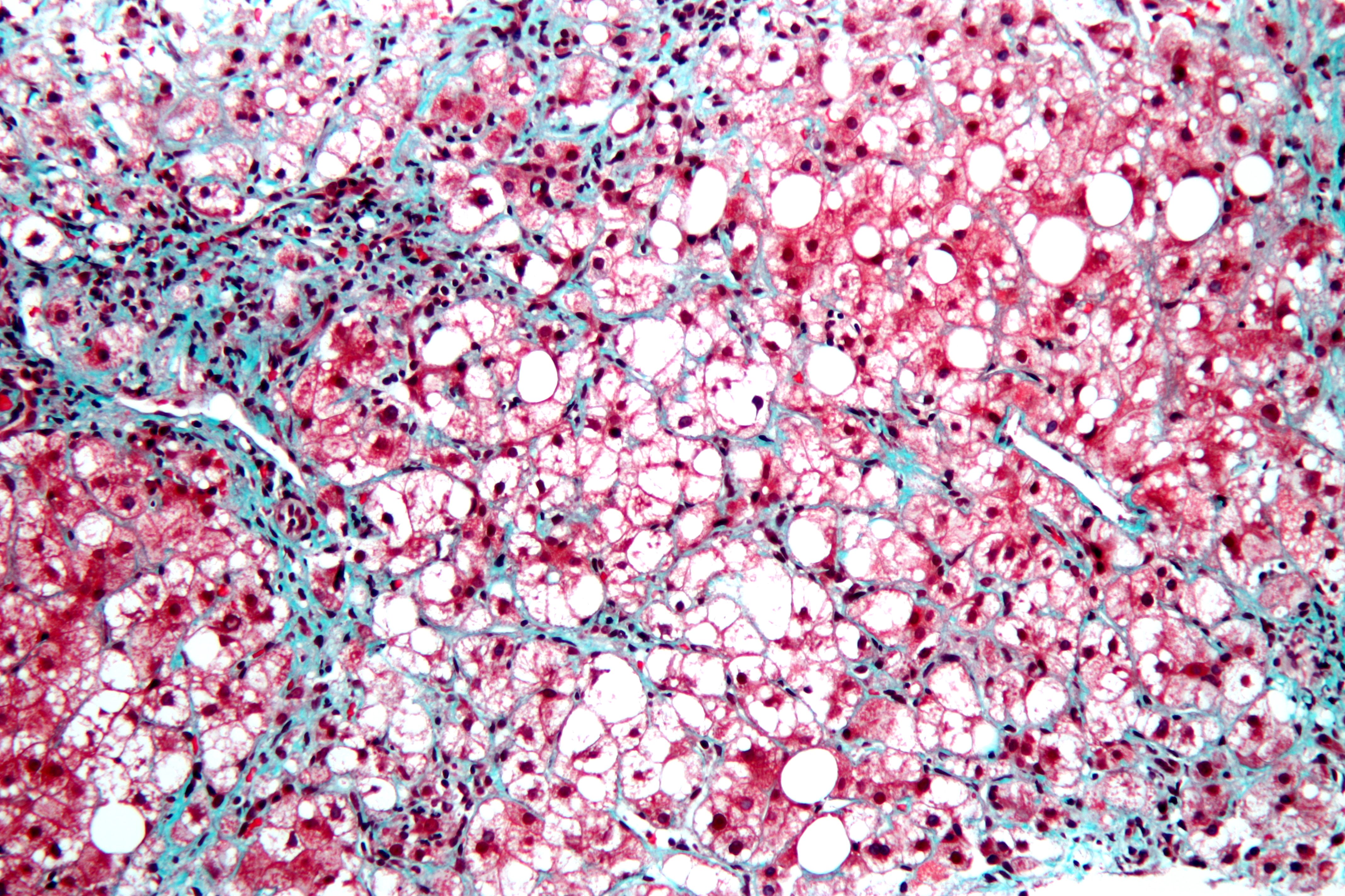
This is a stained liver biopsy sample showing advanced cellular changes in non-alcoholic fatty liver disease. Blue is fibrosis. White is fat accumulation in degenerated cells. Courtesy of Nephron's work.
This is a stained liver biopsy sample showing advanced cellular changes in non-alcoholic fatty liver disease. Blue is fibrosis. White is fat accumulation in degenerated cells. Courtesy of Nephron’s work.

What Is Non-Alcoholic Fatty Liver Disease?

[dropcap]N[/dropcap]on-alcoholic fatty liver is a non-inflammatory liver disorder characterized by degenerative changes in the liver caused by excessive accumulation of lipid (fat) in hepatocytes (liver cells) that is called free fatty acid-generated lipotoxicity.

Non-alcoholic fatty liver shows an increase in liver enzymes called transaminases.

Q: What are the enzymes that increase?

A: The transaminases that increase are ALT and AST. ALT is the abbreviation for alanine aminotransferase enzyme and AST is the abbreviation for aspartate aminotransferase enzyme. They are commonly measured in blood tests to determine liver function and when elevated indicate inflammation.

What Is Non-Alcoholic Fatty Liver In Celiac Disease and/or Gluten Sensitivity?

Hashimoto’s Disease (Autoimmune Thyroiditis Causing Hypothyroidism)

DyspareuniaWhat Is Autoimmune Thyroiditis (Hypothyroidism)?

[dropcap]A[/dropcap]utoimmune thyroiditis, also called Hashimoto’s thyroiditis or Hashimoto’s Disease, is an autoimmune destruction of thyroid tissue characterized by insufficient thyroid hormone circulating in the body that causes formation of a goiter (enlarged thyroid gland) and hypothyroidism.

Hypothroidism refers to the condition of markedly reduced secretion of thyroid hormone. There are other causes of hypothyroidism besides Hashimoto’s thyroiditis.

Hashimoto’s thyroiditis is often associated with other autoimmune diseases such as celiac disease.

In Hashimoto’s thyroiditis, a profusion of antibodies are produced, which build up in the blood. Left untreated so that much of the thyroid gland is destroyed, this condition may progress to the very serious and life-threatening condition called myxedema.

Note: In myxedema, protein, electrolytes, and water abnormally accumulate in between cells which produce firm, inelastic puffy skin that is cool, dry, rough, scaly, and may appear yellow; in some people, areas such as the ankles become crusty with a look of tree bark. Many systemic changes develop shown by significant slowing of mental and physical functions. Please see below.

Q: What thyroid tissue is targeted for destruction?

A: In Hashimoto’s thyroiditis, high levels of autoantibodies target thyroglobulin and thyroid peroxidase, leading to inflammation and destruction of the thyroid gland. The resulting fibrosis or scarring of the gland results in lack of thyroid hormone production.

The thyroid gland consists of a large number of closed vesicles that contain a homogenous substance called colloid, which contains the thyroglobulin. Thyroglobulin is an iodine-containing protein secreted by the thyroid gland and stored within its colloid, from which the thyroid hormones thyroxine (T4) and triiodothyroinine (T3) are derived.1

T3 is the active hormone and is made from T4. Thyroid hormones affect metabolism, brain development, breathing, heart and nervous system functions, body temperature, muscle strength, skin dryness, menstrual cycles, weight, and cholesterol levels.

Thyroid hormone production is regulated by thyroid-stimulating hormone (TSH), which is made by the pituitary gland in the brain. Normally, when thyroid hormone levels in the blood are low, the pituitary releases more TSH. When thyroid hormone levels are high, the pituitary decreases TSH production.

Hashimoto’s disease, with or without the development of hypothyroidism, is treated with synthetic thyroxine, which is man-made T4. Health care providers prefer to use synthetic T4, such as Synthroid® (Levothyroxine), rather than synthetic T3, because T4 stays in the body longer, ensuring a steady supply of thyroid hormone throughout the day. The thyroid preparations made with animal thyroid are not considered as consistent as synthetic thyroid.2

What Is Autoimmune Thyroiditis In Celiac Disease and/or Gluten Sensitivity?

Sources:
  1. Taber’s Cyclopedic Medical Dictionary. 19th ed. F.A. Davis Company. Philadelphia, PA. []
  2. National Endocrine and Metabolic Diseases Information Service. []

Secondary Hyperparathyroidism

Courtesy Wikipedia.com
Parathyroid Glands in the Neck. Courtesy Wikipedia.com

What Is Secondary Hyperparathyroidism?

[dropcap]S[/dropcap]econdary hyperparathyroidism is a parathyroid disorder resulting from hypocalcemia (low blood calcium level) that is characterized by excessive production of parathyroid hormone in the attempt to normalize the low blood calcium by releasing calcium from bone.

Parathyroid hormone is produced by the four pea sized parathyroid glands that are located on the thyroid gland in the front of the neck. In part, because the thyroid and parathyroid glands share the same anatomic place in the body and because they have similar names, they are often confused although they have completely different actions.

Parathyroid hormone regulates calcium and the opposing mineral phosphorus in the blood. In secondary hyperparathyroidism, calcium blood levels are low to normal while phosphorus levels are increased which stimulates the outpouring of parathyroid hormone.

Q: How does secondary hyperparathyroidism differ from primary hyperparathyroidism?

A: In primary hyperparathyroidism blood calcium is high and phosphorus is low, which is the opposite of secondary hyperparathyroidism.

The most common cause of secondary hyperparathyroidism is kidney disease causing failure to reabsorb calcium followed by vitamin D deficiency and malabsorption.

What Is Secondary Hyperparathyroidism In Celiac Disease and/or Gluten Sensitivity?

Selenium Deficiency

selenium gluten free celiac disease symptomWhat Is Selenium?

[dropcap]S[/dropcap]elenium is a mineral that is required by the body  in trace amounts for a healthy immune system, normal thyroid function, and antioxidant protection.

Selenium is absolutely required in the production of at least 30 selenoproteins in the body. First, selenium is joined to the amino acids cysteine as selenocysteine and to methionine as selenomethionine before being used as components for selenoproteins. Many selenoproteins are important antioxidant enzymes such as glutathione peroxidase.

Q: How does glutathione peroxidase work?

A: Glutathione peroxidase activity helps the recycling of vitamins C and E in optimizing the performance of the antioxidant system. The antioxidant properties of selenoproteins help prevent cellular damage from free radicals. Free radicals are natural by-products of oxygen metabolism that may contribute to the development of chronic diseases such as cancer and heart disease.

In the immune system, selenium stimulates immune properties of lymphocytes (white blood cells) by contributing to higher natural killer lymphocyte activity. Natural killer lymphocytes have the ability to destroy cancer cells and bacterial and viral agents.

Other selenoproteins help protect the thyroid gland from anti-oxidants and regulate thyroid function. Specifically, selenium plays an integral role in thyroid gland metabolism.1 Functions are more fully described below.

According to the Food and Agriculture Organization, United Nations, approximately 30 percent of tissue selenium is contained in the liver, 15 percent in kidney, 30 percent in muscle, and 10 percent in blood plasma.

What Is Selenium Deficiency In Celiac Disease and/or Gluten Sensitivity?

Sources:
  1. Stazi AV, Trinti B. Selenium status and over-expression of interleukin-15 in celiac disease and autoimmune thyroid diseases. Ann Ist Super Sanita. 2010;46(4):389-99.DOI: 10.4415/ANN_10_04_06. []

Hypoparathyroidism, Idiopathic

Parathyroid gland anatomyWhat Is Idiopathic Hypoparathyroidism?

[dropcap]I[/dropcap]diopathic hypoparathyroidism is a metabolic condition that results from reduced secretion or impaired action of parathyroid hormone (PTH) which results in a combination of low calcium and elevated phosphorus levels in the body.

Calcium and phosphorus are minerals that act in opposition to each other in the body. Idiopathic means the cause is not known.

Parathyroid hormone is produced by the four pea sized parathyroid glands that are located on the thyroid gland in the front of the neck. Partly because the thyroid and parathyroid glands share the same anatomic place in the body and partly because they have similar names, they are often confused although they have completely different actions.

Q: What does parathyroid hormone do in the body?

A: This vital hormone maintains a correct balance of calcium and phosphorous in the bloodstream and ultimately has an effect on all organs because of the complexity of intracellular calcium physiology.1 

Parathyroid hormone is required to sustain life, therefore undetected or misdiagnosed hypoparathyroidism may pose a significant threat to health outcomes, as its presence may increase disease and mortality in affected individuals.

The clinical consequences of parathyroid hormone deficiency or impaired receptor action involve many body functions. In some patients, however, its manifestation may be non-specific, and in these cases the correct diagnosis may be easily missed.

Digestive manifestations of hypoparathyroidism are few and consist mainly of steatorrhea due to insufficient meal-stimulated cholecystokinin secretion by the duodenal mucosa.2

Laboratory measurements show hypocalcemia (low blood calcium level), hyperphosphatemia (high blood phosphate level), and inappropriately low or undetectable parathyroid hormone levels. Treatment consists of oral calcium supplementation and vitamin D derivatives.3

Idiopathic hypoparathyroidism is rare, although there is a growing incidence of the autoimmune form of hypoparathyroidism, which may occur in combination with other autoimmune diseases such as celiac disease.

What Is Idiopathic Hypoparathyroidism In Celiac Disease and/or Gluten Sensitivity?

Sources:
  1. Abboud B, Daher R, Boujaoude J. Digestive manifestations of parathyroid disorders. World J Gastroenterol. 2011 Sep 28;17(36):4063-6. doi: 10.3748/wjg.v17.i36.4063. []
  2. Abboud B, Daher R, Boujaoude J. Digestive manifestations of parathyroid disorders. World J Gastroenterol. 2011 Sep 28;17(36):4063-6. doi: 10.3748/wjg.v17.i36.4063. []
  3. Krysiak R, Handzlik-Orlik G, Kedzia A, Machnik G, Okopień B. Hypoparathyroidism: the present state of art. Wiad Lek. 2013;66(1):18-29. []

Cancer, Parathyroid

CT Scan of Parathyroid Carcinoma Displacing the Trachea. Courtesy Orphanet Journal of Rare Diseases
CT Scan of Parathyroid Carcinoma Displacing the Trachea by Doina Piciu. Courtesy Orphanet Journal of Rare Diseases

What Is Parathyroid Carcinoma?

[dropcap]P[/dropcap]arathyroid carcinoma is a slow growing rare malignancy involving overactive parathyroid glands and is characterized by profound hypercalcemia (elevated blood calcium level), parathyroid hormone levels of more than 3 times upper normal limits, and palpable neck mass.1

Parathyroid carcinoma is a cause of severe hyperparathyroidism. Hyperparathyroidism results from overproduction of parathyroid hormone by the parathyroid glands.

Research comparing patients with carcinoma to patients with primary hyperparathyroidism showed that patients with parathyroid carcinoma had significantly higher serum parathyroid hormone and calcium levels compared with patients with primary hyperparathyroidism.2

Q: Why is the blood calcium level elevated?

A: Blood calcium level is elevated due to overproduction of parathyroid hormone by the diseased gland(s).

The function of parathyroid hormone (PTH) is to keep calcium blood levels normal, which it does by drawing calcium out of bones as needed. The problem of too much PTH is that bone is constantly robbed of calcium and this depletion causes bone demineralization, resulting in osteopenia progressing to osteoporosis. Of course, weakened bones are subject to breakage with light trauma.

On the other hand, too much calcium in the blood can cause kidney stone formation because urine production is the way the body excretes excess calcium. That is, highly concentrated calcium precipitates out of the urine solution to form stones.

With an estimated incidence of 0.015 per 100,000 population and an estimated prevalence of .005% in the United States, parathyroid cancer is one of the rarest of all human cancers according to the National Cancer Institute.

What Is Parathyroid Carcinoma In Celiac Disease and/or Gluten Sensitivity?

Sources:
  1. Schaapveld M, Jorna FH, Aben KK, Haak HR, Plukker JT, Links TP. Incidence and prognosis of parathyroid gland carcinoma: a population-based study in The Netherlandsestimating the preoperative diagnosis. Am J Surg. 2011 Nov;202(5):590-7. doi: 10.1016/j.amjsurg.2010.09.025. Epub 2011 Aug 20. []
  2. Schaapveld M, Jorna FH, Aben KK, Haak HR, Plukker JT, Links TP. Incidence and prognosis of parathyroid gland carcinoma: a population-based study in The Netherlandsestimating the preoperative diagnosis. Am J Surg. 2011 Nov;202(5):590-7. doi: 10.1016/j.amjsurg.2010.09.025. Epub 2011 Aug 20. []

Autoimmune Hepatitis


Jaundice, or yellowing of the skin and sclera of eyes.
Jaundice, or yellowing of the skin and sclera of eyes, is a symptom of autoimmune hepatitis.

What Is Autoimmune Hepatitis?

[dropcap]A[/dropcap]utoimmune hepatitis is an autoimmune attack against liver cells that is characterized by inflammation and results in chronic liver disease which includes low blood albumin and cirrhosis.

Autoimmune hepatitis is diagnosed by detection of autoantibodies in blood and liver biopsy.

Q: What is cirrhosis?

A: Cirrhosis is a degenerative process of the liver that can be fatal. Normal lobular liver structure is distorted and replaced with nodules of regenerating liver cells separated by bands of fibrous tissue that cannot properly carry out liver function and block the necessary flow of blood through the liver, ending in liver failure.

The liver is the largest organ within the body and lies mostly in the upper part of the abdomen on the right side just under the diaphragm. About 70% of liver tissue is made up of cube shaped cells called hepatocytes that do the main work of the liver. Other cells form structure and are arranged in single layers around blood vessels, sinusoids, and bile ducts.

Bile ducts carry bile, a greenish brown liquid made by the liver, to the gall bladder for storage until needed to aid in the digestion and absorption of fat from the small intestine. Bile emulsifies fat eaten in the diet so that the pancreatic enzyme called lipase can break it down into its fatty acid and glycerol components.

The liver is a very busy organ, carrying out over 500 metabolic processes! Nutritionally, it is the first to receive all the blood carrying nutrients freshly absorbed from the digestive tract. Hepatocytes then convert the nutrients to provide the body with energy, a host of amino acids as building blocks for numerous cell activities, and proteins such as albumin and fibrinogen. In fact, hepatocytes build the structural proteins for the liver itself. Imagine the array of nutrients the liver uses and keeps at its disposal for its metabolic functions. Included in liver storage are vital vitamins and minerals such as vitamin A and vitamin B12.

As a detoxifier, Kupffer cells located in the sinusoids act like amoebas to engulf and digest unwanted matter. As a blood cleanser, the liver removes waste products produced by normal metabolism and toxic substances and rids it by preparing these substances for elimination in urine, such as ammonia converted to urea, and/or excreting them in bile for eventual elimination in stool. Bile is continually made by the liver from phospholipids salt, cholesterol, aging blood cells it removes from circulation.

Autoimmune hepatitis is classified into several types. Type 1 autoimmune hepatitis is the most common form in North America. Type 1 can occur at any age; however, it most often starts in adolescence or young adulthood. People with type 1 autoimmune hepatitis commonly have other autoimmune disorders (see listed below).

Type 2 autoimmune hepatitis is less common and occurs more often in children than adults. People with type 2 can also have any of the autoimmune disorders (see listed below).

Both types of autoimmune hepatitis are treated with prednisone, a corticosteroid hormone to control inflammation and reduce the body’s immune activity against the liver.1

What Is Autoimmune Hepatitis In Celiac Disease and/or Gluten Sensitivity?

Sources:
  1. National Digestive Diseases Information Clearinghouse []

Liver or Hepatic Granulomatous Disease 

Microscopic View of a Liver Granuloma. Courtesy pathology.med.umich.edu/greensonlab.

What Is Hepatic Granulomatous Disease?

[dropcap]H[/dropcap]epatic granulomatous disease is an infiltrative chronic liver disorder characterized by growth of small granulomas.

Rather than being a final diagnosis, the presence of hepatic granulomas entails the need for further investigations towards identifying the underlying etiology, with a pathologist being at the center of the diagnostic process.1

Q: What are hepatic granulomas?

A: Hepatic granulomas are unique inflammatory formations that require liver biopsy for diagnosis.2

Hepatic granulomas develop through the interactions of T lymphocytes and macrophages, with the integral involvement of T-helper (T(H)) 1 or T(H)2 pathways or both, depending on the specific granulomatous disease.

Hepatic granulomas may be detected clinically by elevated levels of serum alkaline phosphatase and g-glutamyltransferase enzymes, damage to specific structures (e.g., intrahepatic bile ducts in primary biliary cirrhosis), or infrequently, progressive liver disease with portal hypertension and cirrhosis (e.g., sarcoidosis).

Systemic immunologic disorders, infectious diseases, drug hepatotoxicity, and reaction to neoplastic disease are the major causative factors responsible for granulomas in the liver.3

What Is Hepatic Granulomatous Disease In Celiac Disease and/or Gluten Sensitivity?

Sources:
  1. Turhan N, Kurt M, Ozderin YO, Kurt OK. Hepatic granulomas: a clinicopathologic analysis of 86 cases. Pathol Res Pract. 2011 Jun 15;207(6):359-65. doi: 10.1016/j.prp.2011.03.003. Epub 2011 Apr 30. []
  2. Bhardwaj SS, Saxena R, Kwo PY. Granulomatous liver disease. Curr Gastroenterol Rep. 2009 Feb;11(1):42-9. []
  3. Lagana SM1, Moreira RK, Lefkowitch JH. Hepatic granulomas: pathogenesis and differential diagnosis. Clin Liver Dis. 2010 Nov;14(4):605-17. doi: 10.1016/j.cld.2010.07.005. []

Diabetes Mellitus, Type I

Microscopic view of pancreatic islet cells. Courtesy Dr. José Sánchez Gonzales

What Is Type I Diabetes Mellitus?

[dropcap]T[/dropcap]ype 1 diabetes mellitus (T1DM), also termed type 1A, is an inherited autoimmune disorder in which anti-islet autoantibodies destroy the islet cells of the pancreas that secrete insulin hormone. Type 1 diabetes mellitus was formerly called juvenile diabetes because it usually afflicts persons under the age of 25 years.

Loss of insulin production results in failure to metabolize glucose. Glucose is a simple sugar that is a required source of energy for the body, especially the brain and muscles.

Type 1 diabetes mellitus is characterized by sustained fasting blood glucose levels above 126 mg/dL (hyperglycemia) with subsequent loss of glucose from the body by removal through the urine (glucosuria) as the body attempts to lower blood glucose, and cell starvation that follows.

That is, while glucose accumulates in blood, the body cannot access it. Without insulin treatment, this disorder quickly produces coma and ultimately results in death. In fact, it is 5th leading cause of death in the United States.

Q: How does insulin work?

A: Insulin moves glucose from the bloodstream into body cells where it is used or reformulated for high energy storage. For example, muscles can use glucose for immediate work or store it in the form of glygogen for later work, depending on need. Healthy insulin production keeps an 8 hour fasting blood glucose level to less than 100 mg/dL. Upon eating carbohydrate food, glucose is digested and absorbed from the small intestine into the bloodstream which then raises blood glucose levels. The elevated level is controlled by prompt action of insulin to lower it to below 140 mg/dL  within 2 hours of eating.

Insulin does not work alone. The islets of Langerhans manage glucose in the body. The islets are specialized formations located on the outer surface of the pancreas. The islets are composed of two different types of cells known as alpha and beta cells. These cells make the competing hormones that keep blood glucose within a healthy range.

Alpha cells secrete glucagon to raise blood glucose levels by triggering the body to release stored energy in the form of glycogen. In the opposite, beta cells secrete insulin to lower blood glucose by opening body cells so that glucose in blood can enter. Without insulin, glucose cannot enter cells but remains in the bloodstream where it accumulates.

Insulin is also needed to move magnesium into cells from the bloodstream. On the other side, magnesium is needed to produce insulin. Insulin has other functions such as building muscle and helping regulate cholesterol which directly impacts the sex hormones, estrogen, progesterone, and testosterone.

Onset of symptoms usually occurs over a period of days or weeks, although beta cell destruction can begin years earlier. The SEARCH for Diabetes in Youth multicenter study, funded by the Centers for Disease Control and Prevention (CDC) and the National Institutes of Health (NIH), has determined that based on data from 2002 to 2003, a total of 15,000 youth in the United States were newly diagnosed with type 1 diabetes each year. Non-Hispanic white youth had the highest rate of new cases of type 1 diabetes according to NIH.

Type 1A diabetes mellitus has become one of the most intensively studied autoimmune disorders. It is now possible to predict its development, beginning with HLA-encoded genetic susceptibility, followed by the development of a series of anti-islet autoantibodies.1

What Is Type I Diabetes Mellitus In Celiac Disease and/or Gluten Sensitivity?

Sources:
  1. Liu E, Eisenbarth GS. Type 1A diabetes mellitus-associated autoimmunity. Endocrinology and Metabolism Clinics of North America. Jun 2002;31(2):391-410, vii-viii. []

Diabetic Instability 

Glucose Meter Reading Showing Hypoglycemia. Courtesy Abbott.
Glucose Meter Reading.Courtesy Abbott.

What Is Diabetic Instability?

[dropcap]D[/dropcap]iabetic instability, also called brittle diabetes, is poor control of blood sugar characterized by frequent rise (hyperglycemia) and fall (hypoglycemia). These fluctuations can be life-threatening and cause unnecessary complications.

Q: What are complications in diabetes?

A: Complications of diabetes include any/all of the following:

  • Peripheral neuropathy that causes burning, numbness, or tingling in the extremities.
  • Skin infections and ulcers that do not heal and can lead to amputation of toes, feet or legs.
  • Damage to retina that can progress to blindness.
  • Cataracts and glaucoma that impair vision and can cause blindness.
  • Increased susceptibility to serious infections of the genitourinary tract, lung and upper respiratory tract that can become systemic and life-threatening.
  • Kidney disease that can lead to kidney failure.
  • Heart disease that can lead to heart failure.
  • Vascular disease that can lead to stroke.
  • Impaired motility in digestive tract that can lead to dyspepsia and constipation.
  • Pregnancy complications.
  • Shortened life expectancy.

The main causes of brittleness include malabsorption, certain drugs (alcohol, antipsychotics), defective insulin absorption or degradation, defect of hyperglycemic hormones especially glucocorticoid and glucagon, and above all delayed gastric emptying as a result of autonomic neuropathy.1

What Is Diabetic Instability In Celiac Disease and/or Gluten Sensitivity?

Sources:
  1. Vantyghem MC, Press M. Management strategies for brittle diabetes. Ann Endocrinol (Paris). 2006 Sep;67(4):287-96. []