Skip to content

Sucrose Intolerance and Sucrosemia 

Sweet Bon Bons: Courtesy Freepik.
Sugary Bon Bons: Courtesy Freepik.

What Is Sucrose Intolerance And Sucrosemia?

[dropcap]S[/dropcap]ucrose intolerance is the inability to digest sucrose, a widely available sugar, while sucrosemia is the abnormal presence of sucrose in the bloodstream.

Q: Why cannot the body digest sucrose?

A: Sucrose, such as cane or beet sugar, is a double molecule sugar which must first be digested before being absorbed from the gut into the bloodstream. That is, sucrose must be split into its component single molecules of fructose and glucose, which are then properly absorbed.

The inability to properly digest sucrose results directly from low production and activity of sucrase in the small intestine. Sucrase is the specific enzyme that splits or digests sucrose.

Undigested sucrose does not remain idle. Its presence acts osmotically to draw water from the body into the intestine, causing watery diarrhea.

Meanwhile, microbiota (normal bacteria) in the colon eagerly ferment the abnormally present sucrose that arrives from the small intestine. Fermentation generates short-chain fatty acids and hydrogen gas, which results in bloating pain.1

In sucrosemia, sucrose molecules abnormaly pass through an unhealthy small intestinal lining and enter the bloodstream where there presence is abnormal. Sucrose in the blood is filtered out by the kidneys and excreted in urine.

Positive response to a breath hydrogen test (BHT), involving 1 – 3 hours of time post ingestion of sucrose test dose, signifies malabsorption in the small intestine and fermentation in the colon. If BHT is positive before 60 minutes, the result implies bacteria is abnormally present in the small intestine, causing fermentation there. Endoscopy is used to measure sucrase activity in tissue samples.

What Is Sucrose Intolerance And Sucrosemia In Celiac Disease and/or Gluten Sensitivity?

Sources:
  1. Krause’s Food, Nutrition, & Diet Therapy. 10th Edition. Kathleen Mahan, Sylvia Escott-Stump. 2000. W.B. Saunders Company. []

Vomiting

VomitingWhat Is Vomiting?

[dropcap]V[/dropcap]omiting is the regurgitation of swallowed food back out of the mouth.

What Is Vomiting In Celiac Disease and/or Gluten Sensitivity?

Tongue – Beefy Red, Smooth, Burning 

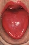
tongue in B12 deficiencyWhat Is Beefy Red, Smooth, Burning Tongue?

[dropcap]B[/dropcap]eefy, red, smooth, burning tongue is an inflammatory alteration in tongue tissue characteristic of vitamin B12 deficiency.

Inflammation makes the tongue red and sore which, when untreated, progresses to atrophied papillae (shrunken taste buds) that makes the tongue smooth.

Q: Why does vitamin B12 alter the tongue?

A: Vitamin B12 is essential for the metabolism of all cells, especially for those of the digestive tract of which the tongue is an organ.1

These easily seen changes to tongue tissue are an important diagnostic feature of deficient vitamin B12, which causes other devastating unseen damage, because the discomfort generally causes the individual to seek medical care.

What Is Beefy Red, Smooth, Burning Tongue In Celiac Disease and/or Gluten Sensitivity?

Sources:
  1. Krause’s Food, Nutrition, & Diet Therapy. 10th Edition. Kathleen Mahan, Sylvia Escott-Stump. 2000. W.B. Saunders Company. []

Tongue – Fiery Red, Smooth, Burning

niacin deficiencyWhat Is A Fiery Red, Smooth, Burning Tongue?

[dropcap]A[/dropcap] fiery red, smooth, burning tongue is an alteration in tongue tissue that is characteristic of advanced niacin deficiency.1

Q: How does niacin deficiency cause the tongue to be red and sore?

A: Niacin is an essential B vitamin that is required for a healthy tongue and by all body cells as well. Deficiency first shows in tissues with rapid cellular turnover, such as mucosal cells of the tongue.

When absorbed from the small intestines, niacin (the form in food) becomes the active form niacinamide. Niacinamide is converted by the body into co-enzymes which are present in all cells. These enzymes function in oxidation-reduction reactions essential for release of energy from carbohydrates, fats, and proteins and are needed as components for more than 200 enzymes involved in metabolism.

In addition to producing energy, niacinamide is essential for healthy skin and the mucosal lining of the digestive tract, normal functioning of the brain and nervous system, and production of steroid hormones from adrenal glands and hormones from sex glands.

What Is A Fiery Red, Smooth, Burning Tongue In Celiac Disease and/or Gluten Sensitivity?

Sources:
  1. Krause’s Food, Nutrition, & Diet Therapy. 10th Edition. Kathleen Mahan, Sylvia Escott-Stump. 2000. W.B. Saunders Company. []

Food Allergy – IgE and Non IgE

Baby with Allergic Reaction to Peanuts. GFW
Baby with Allergic Reaction to Peanuts. GFW photo.

What Is Food Allergy?

[dropcap]F[/dropcap]ood allergy is an abnormal immune response to food proteins that may damage the small intestinal  lining and produce malabsorption of food. The reaction is usually delayed which makes it difficult to identify the offending food that is causing symptoms.

Q: How does food allergy develop?

A: The gastrointestinal tract serves not only to digest and absorb foodstuffs but also to protect the body from unwanted substances. When allergic food substances are eaten, the immune response that is triggered in the gut produces inflammation with symptoms such as pain, vomiting and loose bowels.

Inflammation causes swelling of the gut lining that can interfere with the passage of nutrients through it to the body which results in malabsorption. Malabsorption deprives the body of nutrients needed for normal function.

Symptoms other than digestive may involve skin rashes, hives, and respiratory difficulties that can be distressing and life-threatening.

What Is Food Allergy In Celiac Disease and/or Gluten Sensitivity?

Vitamin B2 (Riboflavin) Deficiency

Chicken Kabobs on the Grill...umm Riboflavin.
Chicken Kabobs on the Grill…umm Riboflavin.

What Is Riboflavin?

[dropcap]R[/dropcap]iboflavin, also called vitamin B2, is fundamentally required for the breakdown of carbohydrates, amino acids, and fats in metabolism, enables oxygen to be used by cells, and acts as a component of more than a dozen enzymes involved in energy production and use.

Some enzymes needing riboflavin also require magnesium to properly function.

In other activities, riboflavin is required for healthy cardiovascular and nervous systems, eye health, and supports antioxidant protection.  Functions are more fully described below.

Urinary excretion of riboflavin cannot be detected when vitamin intake is below the required levels. On the other hand, when intake exceeds saturation in the body, the vitamin and/or its metabolites are actively excreted into urine to prevent excessive toxicity of the vitamins.1

There is no specific name for riboflavin deficiency disease.

What Is Riboflavin Deficiency In Celiac Disease and/or Gluten Sensitivity?

Sources:
  1. Shibata K, Hirose J, Fukuwatari T. Relationship Between Urinary Concentrations of Nine Water-soluble Vitamins and their Vitamin Intakes in Japanese Adult Males. Nutr Metab Insights. 2014 Aug 5;7:61-75. doi: 10.4137/NMI.S17245. []

Plummer-Vinson Syndrome Affecting Esophagus

mucosal web in plummer-vinson syndromeWhat Is Plummer-Vinson Syndrome Affecting the Esophagus?

[dropcap]P[/dropcap]lummer-Vinson syndrome is a manifestation of severe, long-term, iron deficiency anemia that is characterized by post-cricoid esophageal webs and dysphagia.

Q: What are esophageal webs?

A: Esophageal webs are one or more thin horizontal membranes consisting of squamous epithelium (cells that line the surface of the esophagus) and submucosa. They usually protrude from the anterior (front) wall, extending laterally across the inside esophagus but not to the posterior (rear) wall, which means that they rarely encircle the lumen.1

Dysphagia, or difficulty swallowing, from these webs is commonly painless and intermittent or progressive and may cause obstruction.

Webs can be detected by barium swallow X-ray, but the best way for demonstration is videofluoroscopy and by upper gastrointestinal endoscopy. They appear smooth, thin, and gray with eccentric or central lumen space. The webs typically occur in the upper part of the esophagus and may be missed and accidentally ruptured unless the endoscope is introduced under direct visualization.1

Iron deficiency is believed to decrease the contraction amplitude or force of the esophageal muscle resulting in motility impairment. Slower transit times have been recorded at the proximal and middle parts of the esophagus of Plummer-Vinson syndrome patients compared to healthy volunteers.2 Transit time is how fast ingested food and fluids travel through the esophagus.

Gude et al, report that iron replacement does not necessarily reverse the dysphagia in all the cases of Plummer-Vinson syndrome and that close monitoring of the web is mandated to watch for malignant change.  In fact, 3 to 15 per cent of the patients with Plummer-Vinson syndrome, mostly women between 15 and 50 years of age, have been reported to develop esophageal or pharyngeal cancer.2

What Is Plummer-Vinson Syndrome Affecting the Esophagus In Celiac Disease and/or Gluten Sensitivity?

Sources:
  1. Novacek G. Plummer-Vinson syndrome. Orphanet J Rare Dis. 2006; 1: 36. Published online 2006 September 15. doi:  10.1186/1750-1172-1-36. [] []
  2. Gude D, Bansal DP, and Malu A. Revisiting Plummer Vinson Syndrome. Annals of Medical and Health Sciences Research. 2013 Jan-Mar;3(1):119-121. [] []

Constipation Alternating with Diarrhea

Bristol Stool Chart Showing Normal and Abnormal Stool.

What Is Chronic Constipation Alternating With Diarrhea?

[dropcap]C[/dropcap]hronic constipation alternating with diarrhea is an intestinal motility disorder, or irregularity, characterized by alteration in stool formation, consistency, and evacuation which results in a bowel movement that consists of some hard or balled stool along with some loose stool that can cause leakage.

Q: How do irregular movement patterns develop in the colon?

A: The colon produces irregular movements as a result of problems that originate in  the colon (large intestine) itself and/or the small intestine which then affects function of the colon.

Here are listed the many types of problems or diseases that cause these abnormal bowel movements:

  • Disorders that adversely affect the colon, an organ which must propel stool, remove excess water, absorb electrolytes, ferment undigested food material that passes into it, and produce nutrients from the fermentation process:
  • Poor diet that does not contain adequate nutrition, fiber, probiotics, prebiotics, and water to form normal stool.
  • Diet that contains irritating, toxic or allergenic food that cause spasms.
  • Diseases that inflame the mucosa lining such as collagenous colitis, altering the proper absorption of water and electrolytes.
  • Diseases that damage and swell the colon walls, such as Crohn’s disease, ulcerative colitis, and diverticulitis.
  • Diseases that obstruct the lumen or passageway so that stool passes with difficulty.
  • Diseases that hamper normal peristalsis (muscle action), such as irritable bowel syndrome (IBS), diabetes and thyroid disease.
  • Disorders that adversely affect the small intestine, an organ which must digest and absorb nutrients needed by the body while passing unabsorbed food material to the colon:  
  • Diet that conatins too much fat, sugar or artifical sweeteners, causing diarrhea.
  • Disorders that result in malabsorption, such as gluten enteropathy, milk enteropathy, steatorrhea (fat malabsorption), lactose intolerance, sucrose intolerance, maltose intolerance, and bacterial overgrowth, passing abnormal amounts of undigested food material to the colon where it is fermented producing excessive gas, diarrhea and spasm.
  • Disorders that impair peristalsis, such as active celiac disease, diabetes, scleroderma, and thyroid disease.
  • Tumors like cancer and lymphoma impair regular passage of material to colon.
  • Drugs that impair peristalsis, such as iron supplements, aluminum containing antacids, narcotics, some anti-depressants, some anti-seizure, and some diuretics.

What Is Chronic Constipation Alternating With Diarrhea In Celiac Disease and/or Gluten Sensitivity?

Bleeding, Occult Gastrointestinal

Erosions Of The Esophagus Can Be A Cause Of Gastrointestinal Bleeding.
Erosions Resulting From Esophagitis Can Be A Cause Of Gastrointestinal Bleeding.

What Is Occult Gastrointestinal Bleeding?

[dropcap]O[/dropcap]ccult gastrointestinal bleeding is characterized by unseen or minute quantities of blood in stool. The origin of bleeding is from mucosa that lines the inside of the digestive tract at a site that must be found by medical testing and procedures to look at the lining.

Q: What tests and procedures are performed to locate the bleeding?

A: The simplest test to discover blood that cannot be seen is the stool test. This consists of a card with 3 spaces for 3 separate  stool collection over 3 separate days. A tiny smear of stool is placed on a test card space on each of three days. Then the card is examined for a reaction that indicates the existence of blood in the stool.

If the stool test is positive, the origin of bleeding must be located. This search requires certain procedures that look at the mucosal lining directly by a gastroenterologist, usually under light sedation.

  • Gastroscopy procedure can visualize the upper gastrointestinal tract, which includes the esophagus and stomach.
  • Endoscopy procedure can also visualize the upper small intestine. However, endoscopy procedure is limited in that it cannot visualize the middle small intestine (jejunum).
  • Colonoscopy can visualize the end of the small intestine and colon all the way to the rectum.
  • Capsule endoscopy procedure.  If  gastroendoscopy and colonoscopy fail to discover the source of bleeding, the physician may administer a capsule endoscopy which visualizes the entire gastrointestinal tract. In this procedure, the patient swallows a capsule size camera (encased) which takes constant pictures over the course of a day until expelled through the rectum.  Of course, the patient must be able to swallow the large size capsule.

What Is Occult Gastrointestinal Bleeding In Celiac Disease and/or Gluten Sensitivity?

Cirrhosis, Primary Biliary

biliary system primary biliary cirrhosisWhat Is Primary Biliary Cirrhosis?

[dropcap]P[/dropcap]rimary biliary cirrhosis (PBC) is a biliary tract disease characterized by chronic cholestasis (build-up of bile) and gradual destruction of bile ducts within the liver, called intrahepatic bile ducts, caused by chronic inflammation.

Primary biliary cirrhosis comes under the umbrella term autoimune liver disease in which the end result is immune-mediated hepatocellular (liver cell) or hepatobiliary (bile duct) injury.1

Q: What is the end result of destruction of bile ducts?

A: The end result of destruction of bile ducts is liver damage.

Injured liver tissue from chronic inflammation and the buildup of bile leads to cirrhosis, a condition in which the liver slowly deteriorates and malfunctions.

Scar tissue replaces healthy liver tissue, partially blocking the flow of blood through the liver. Scarring also impairs the liver’s normal ability to control infections, remove bacteria and toxins from the blood, process nutrients, hormones, and drugs, make proteins that regulate blood clotting, produce bile, and effectively replace its own cells when they become damaged.2

The liver is the largest organ within the body. It lies mostly in the upper part of the abdomen on the right side just under the diaphragm. About 70% of liver tissue is made up of cube shaped cells called hepatocytes that do the main work of the liver. Other cells (epithelial) form structure and are arranged in single layers around blood vessels, sinusoids, and bile ducts.

Bile ducts carry bile, a greenish brown liquid made by the liver to the gall bladder for storage until needed to aid in the digestion and absorption of fat and fat-soluble vitamins A, D, E, and K from the small intestine. Bile emulsifies fat eaten in the diet so that the pancreatic enzyme called lypase can break it down into its fatty acid and glycerol components which can then be absorbed into the body.

Bile also carries away waste products produced by normal metabolism and toxic substances that are removed by the liver for eventual elimination in stool. Bile is continually made by the liver from phospholipids, bile acids, cholesterol, and aging blood cells it removes from circulation. As such, bile must continually flow out of the liver to prevent build-up in the liver.

There is no cure for primary biliary cirrhosis.

What Is Primary Biliary Cirrhosis In Celiac Disease and/or Gluten Sensitivity?

Sources:
  1. Trivedi PJ, Adams DH. Mucosal immunity in liver autoimmunity: a comprehensive review. J Autoimmun. 2013 Oct;46:97-111. doi: 10.1016/j.jaut.2013.06.013. []
  2. http://digestive.niddk.nih.gov/ddiseases/pubs/primarybiliarycirrhosis/ []