Skip to content

Systemic Lupus Erythematosus 
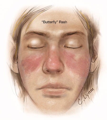
Image showing butterfly rash of SLE. Courtesy JAMA.
Image showing butterfly rash typical of SLE. Courtesy JAMA.

What Is Systemic Lupus Erythematosus?

[dropcap]S[/dropcap]ystemic lupus erythematosus (SLE) is a chronic autoimmune inflammatory disease that is characterized by involvement of multiple organs due to the production of antibodies to components of the cell nucleus.1 SLE has an unpredictable course of acute flare-ups and remissions.

Severity depends on the extent of organs affected with skin and nail involvement, called discoid lupus, being the least serious and inflammmation of the kidney, called lupus nephritis, being the worst.

Nevetheless, a classic presentation is development of a rash over the cheeks and nose that resembles a butterfy with wings spread hence the name “butterfly rash.”

Symptoms are many and varied depending on the tissues affected and are often not specific, for example hair loss has a variety of causes. Symptoms can be confused by co-existence with other autoimmune disease such as Sjogren’s syndrome.

Systemic lupus erythematosus should be managed by a specialist. Symptoms can be controlled with steroid therapy, but this disease can be a cause of premature death  mainly from active disease, organ failure (e.g., kidneys), infection, or cardiovascular disease from accelerated atherosclerosis.

Certain common medicines known to cause drug-induced lupus are:

  • Isoniazid
  • Hydralazine
  • Procainamide

Other less common drugs may also cause the condition. These may include:

  • Anti-seizure medications
  • Capoten
  • Chlorpromazine
  • Etanercept
  • Infliximab
  • Methyldopa
  • Minocycline
  • Penicillamine
  • Quinidine
  • Sulfasalazine

Symptoms tend to occur after taking the drug for at least 3 to 6 months.2

Although there is a strong familial aggregation, the disease is relatively uncommon and most cases are sporadic.1 According to the Center for Diseases (CDC), lupus most commonly affects women of childbearing age but also occurs in infants, children, adolescents, and men with peak occurrence between ages 15 and 40. Blacks (and possibly Hispanics, Asians, and Native Americans) are affected more than Whites.

What Is Systemic Lupus Erythematosus In Celiac Disease and/or Gluten Sensitivity?

Sources:
  1. http://www.cdc.gov/arthritis/basics/lupus.htm [] []
  2. www.nlm.nih.gov/medlineplus/ency/article/000446.htm []

DHA (Docosahexaenoic Acid) Deficiency

Smoked Salmon...Brain Food that's Good for the Eyes!
Salmon…Brain Food That’s Good for Your Eyes, Heart, Blood Vessels, Blood Sugar, Liver, Muscles, and Fights Inflammation!

What Is DHA?

[dropcap]D[/dropcap]ocosahexaenoic acid (DHA) is an essential omega-3 fatty acid that is abundant in the brain, being crucial in brain structure. As such DHA is a key component of neuronal membranes together with arachidonic acid (a major opposing omega-6 fatty acid), making up 15-20% of the brain’s dry mass.

This polyunsaturated fatty acid is obtained from fish sources of food.

In healthy human volunteers, positron emission tomography (PET) has shown that the normal human brain consumes 4.6 mg/day of DHA.1

DHA is particularly concentrated in highly active membranes such as nerve synapses (junctions) and photoreceptors in the eye (retina).

Q: How much DHA is in the retina?

A: DHA makes up more than 30% of the retina.2

In other roles, DHA is an  important building material for the eicosanoids, a large group of highly bioactive hormone-like substances including prostaglandins, leukotrienes, and thromboxanes that are involved in blood clotting, inflammation, and vasoconstriction.

DHA has been shown to increase insulin sensitivity as opposed to the opposite problem of insulin resistance, to improve muscle mass, and protect against non-alcoholic fatty liver disease.3

Egert et al. in a study of people aged 19 to 43 years with normal cholesterol showed that DHA intake significantly increased serum HDL (good) cholesterol. Also, DHA significantly decreased fasting serum triglycerides.4

What Is DHA Deficiency In Celiac Disease and/or Gluten Sensitivity?

Sources:
  1. Rapoport SI. Brain arachidonic and docosahexaenoic acid cascades are selectively altered by drugs, diet and disease. Prostaglandins Leukot Essent Fatty Acids. 2008 Sep-Nov;79(3-5):153-6. Epub 2008 Oct 29. []
  2. Richardson AJ. The importance of omega-3 fatty acids for behavior, cognition, and mood. Scandinavian Journal of Nutrition. 2003;47(2):92-8 []
  3. Espinosa A, Valenzuela R, González-Mañán D, D’Espessailles A, Guillermo Gormaz J, Barrera C, Tapia G. Prevention of liver steatosis through fish oil supplementation: correlation of oxidative stress with insulin resistance and liver fatty acid content. Arch Latinoam Nutr. 2013 Mar;63(1):29-36. []
  4. Egert S, Kannenberg F, Somoza V, et al. Dietary alpha-linolenic acid, EPA, and DHA have differential effects on LDL fatty acid composition but similar effects on serum lipid profiles in normolipidemic humans. J Nutr. 2009;139:861–868. doi: 10.3945/jn.108.103861 []

Homocysteine, Elevated Blood Level (Hyperhomocysteinemia)

A 3-D model of homocysteine.

What Is Elevated Homocysteine?

[dropcap]E[/dropcap]levated homocysteine in blood, called hyperhomocysteinemia, indicates an abnormal blood level of this transient amino acid.

Q: How does the level of homocysteine become abnormal?

A: In metabolism, homocysteine is briefly formed in the breakdown of the amino acid methionine. It is normally converted to cystathione and then to the amino acid cysteine by means of an enzyme that requires vitamin B6.

In the reverse, conversion of homocysteine to methionine requires an enzyme dependent on adequate folic acid and vitamin B12 levels.

Insufficient methionine levels and/or inefficiency in this process results in elevated homocysteine plasma levels that are toxic to blood vessels.

Folic acid, vitamin B12 and vitamin B6 are involved in the metabolic removal of homocysteine, but folic acid deficit occurs the most often.1

What Is Elevated Homocysteine In Celiac Disease and/or Gluten Sensitivity?

Sources:
  1. Lim PO, Tzemos N, Farquharson CA, et al. Reversible hypertension following coeliac disease treatment: the role of moderate hyperhomocysteinaemia and vascular endothelial dysfunction. Journal of Human Hypertension. Jun 2002;16(6):411-5. []

Vasculitis, Cerebral (Cause of Stroke, TIA, and Seizure)

How Vasculitis Develops. Courtesy quizlet.com

What Is Cerebral Vasculitis?

[dropcap]C[/dropcap]erebral vasculitis, also called vasculitis of the central nervous system (CNS), is an autoimune attack against elastin fibers in the walls of arteries that bring blood to the head. Early recogniton may reduce poor outcomes.1

Cerebral vasculitis is characterized by inflammation of large, medium, or small blood vessels which is often segmental with scattered foci (sites) of intense inflammation, and results in necrosis (death) of affected tissues with scarring that occludes, or blocks, blood flow.

Q: What happens when an artery is occluded by scarring?

A: When an artery is occluded by scarring, blood cannot flow through it thus preventing the body tissues it supplies with oxygen and nutrition.   Depending on vessels that are affected, blindness, TIA (transient ischemic attack) or stroke may result from blockage or rupture (hemorrhage).

Blood flow through arteries can be seen by angiography procedure. The diagnosis is made by biopsy. Additionally, contrast-enhanced MRI, proven to be sensitive to extradural arteritis, for the identification of intracranial vessel wall inflammation shows that wall thickening and intramural contrast uptake are frequent findings in patients with active cerebral vasculitis affecting large brain arteries.2

Vasculitis may develop with  autoimmune diseases, such as celiac disease, lupus eythematosis and rheumatoid arthritis due to immune complexing, and possibly severe infection and drug sensitivity.

What Is Cerebral Vasculitis In Celiac Disease and/or Gluten Sensitivity?

Sources:
  1. Salvarani C, Brown RD Jr, Calamia KT, Christianson TJ, Weigand SD, Miller DV, Giannini C, Meschia JF, Huston J 3rd, Hunder GG. Primary central nervous system vasculitis: analysis of 101 patients. Ann Neurol. 2007 Nov;62(5):442-51. []
  2. Küker W, Gaertner S, Nagele T, Dopfer C, Schoning M, Fiehler J, Rothwell PM, Herrlinger U. Vessel wall contrast enhancement: a diagnostic sign of cerebral vasculitis. Cerebrovasc Dis. 2008;26(1):23-9. doi: 10.1159/000135649. Epub 2008 May 30. []