Skip to content

Alopecia, Diffuse (Balding Hair Loss)

Courtesy of slideshow.net

What Is Transient Erythroblastopenia?

T ransient erythroblastopenia is a rare disorder of red blood cell formation characterized by brief, reversible disappearance of erythroblasts (red blood cell precursors) in the bone marrow of children.

Q: What do the red blood cells look like?

A: Circulating red blood cells appear normal so that the anemia which develops is a normocytic (normal cellular) anemia in contrast to microcytic (small cell) anemia in iron deficiency or macrocytic (large cell) anemia in vitamin B12 or folic acid deficiency.

Because new red blood cells are failing to mature, packed red blood cell transfusions may be necessary in severe anemia.

In adults, this disorder is called pure red blood cell anemia.

What Is Transient Erythroblastopenia In Celiac Disease and/or Gluten Sensitivity?

Short Duration of Breast Feeding 

dermatomyositisWhat Is Dermatomyositis?

D ermatomyositis is a rare autoimmune systemic disease of the connective tissue that is characterized by inflammatory and debilitating degenerative changes in the muscles and in the skin. 

Dermatomyositis results in symmetric, proximal muscle weakness of limbs (upper arms and legs), and skin manifestations. 50-70% of patients have circulating myositis-specific auto-antibodies.

The course of dermatomyositis is unpredictable being marked by spontaneous flare-ups and remissions. It can begin slowly or abruptly according to the factor that is triggering the onset such as infection, medications like phenytoin, and autoimmune disease.

Q: What are the skin manifestations of dermatomyositis?

A: Classic skin manifestations of dermatomyositis include these features:

  • The heliotrope rash (lilac color) on upper eyelids.
  • Rash on face, neck, shoulders, upper chest, elbows, knees, knuckles, and back.
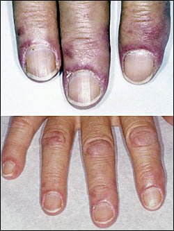
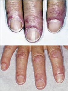
  • Gottron’s papules (scaly, red eruptions or  patches over the knuckles, elbows, and knees).
  • The V-sign (rash front of neck and chest).
  • The shawl sign (rash distribution on shoulders and back).1

Additional cutaneous manifestations are described below under symptoms.

Dermatomyositis is associated with an increased risk of  cancer, other autoimmune diseases, such as lupus and psoriasis, and it can be a complication of interferon-α therapy. About 1 person in 100,000 are affected according to various studies. While it affects all ages, women have twice the occurence of men.

There is no cure for dermatomyositis, but the symptoms can be treated.  Options include medication, physical therapy, exercise, heat therapy (including microwave and ultrasound), orthotics and assistive devices, and rest.  The standard treatment for dermatomyositis is a corticosteroid drug, given either in pill form or intravenously.  Immunosuppressant drugs, such as azathioprine and methotrexate, may reduce inflammation in people who do not respond well to prednisone.2

What Is Dermatomyositis In Celiac Disease and/or Gluten Sensitivity?

Sources:

  1. Marvi U, Chung L, Fiorentino DF. Clinical presentation and evaluation of dermatomyositis. Indian J Dermatol. 2012 Sep;57(5):375-81. doi: 10.4103/0019-5154.100486. []
  2. National Institute of Neurological Disorders and Stroke. []

Attention Deficit Hyperactive Disorder (ADHD)

A small intestinal mucosa from a case of refractory coeliac disease immunostained sequentially for CD3 (alkaline phosphatase-blue) and CD8 (peroxidase-brown). Most intraepithelial lymphocytes are CD3+, CD8-.Courtesy pubcan.org
Mucosa  in refractory celiac  disease immunostained sequentially for CD3 (alkaline phosphatase-blue) and CD8 (peroxidase-brown). Most intraepithelial lymphocytes are CD3+, CD8-. Courtesy pubcan.org

What Is Refractory Celiac Disease?

R efractory celiac disease, formerly called refractory sprue, is a severe complication characterized by persistence of symptoms and intestinal inflammation despite gluten free diet after 12 months.1

Refractory celiac disease appears in two forms, ulcerative jejunitis (RCD I) and cryptic intestinal T-cell lymphoma (RCD II).

Patients with RCD I seem to profit from immunosuppressive treatment, but positive response to corticosteroid treatment does not exclude underlying enteropathy–associated T-cell lymphoma (EATL).

Patients with RCD II have a high percentage of abberant T-cells and is usually resistant to medical therapies. The presence of an aberrant clonal intraepithelial T-cell population has led to the designation of refractory celiac disease with this population as a cryptic intestinal T-cell lymphoma, characterized by frequent dissemination to the blood and the entire gastrointestinal lining.2

  • Refractory sprue may occur after an initial response to gluten free diet or without any evidence of preexisting celiac disease. All other causes of malabsorption must be excluded, such as collagenous colitis.
  • In a subgroup of patients with enteropathy-associated T-cell lymphoma (EATL) there is progressive deterioration of a refractory form of celiac disease. The prognosis is poor, although some patients respond to corticosteroids and immunosuppressive agents.3
  • A nationwide Finnish study showed that patients of male gender, older age, severe symptoms or seronegativity (negative antibody result) at the diagnosis of celiac disease are at risk of future refractory coeliac disease and should be followed up carefully.4
  • Chorea has been described as a paraneoplastic phenomenon in patients with non-Hodgkin’s lymphoma and has been described as associated with lymphoma arising from a background of refractory celiac disease. The finding of chorea in association with celiac disease should prompt a search for possible underlying intestinal T-cell lymphoma.5

How Prevalent Is Refractory Celiac Disease?

Sources:
  1. Murray JA, The widening spectrum of celiac disease. American Journal of Clinical Nutrition. Mar 1999;69 (3):354-365. []
  2. Culliford AN, Green PH. Refractory sprue. Current Gastroenterology Reports. Oct 2003;5(5):373-8. []
  3. Culliford AN, Green PH. Refractory sprue. Current Gastroenterology Reports. Oct 2003;5(5):373-8. []
  4. Ilus T, Kaukinen K, Virta LJ, Huhtala H, Mäki M, Kurppa K, Heikkinen M, Heikura M, Hirsi E, Jantunen K, Moilanen V, Nielsen C, Puhto M, Pölkki H, Vihriälä I, Collin P. Refractory coeliac disease in a country with a high prevalence of clinically-diagnosed coeliac disease. Aliment Pharmacol Ther. 2014 Feb;39(4):418-25. doi: 10.1111/apt.12606. []
  5. Kitiyakara T, Jackson M, Gorard DA. Refractory coeliac disease, small-bowel lymphoma and chorea. J R Soc Med. 2002 Mar;95(3):133-4. []

Hypocalcemia (Low Blood Calcium)

hypocalcemia celiac disease gluten symptomWhat Is Hypocalcemia?

Hypocalcemia, or low plasma calcium, means the level of calcium in blood is too low to meet metabolic needs of the body for calcium.

Low blood calcium is characterized by bone and tooth demineralization (loss of calcium causing weak teeth and fragile bones), and these impaired functions: nerve conduction, muscle contraction, blood clotting, blood pressure regulation, glycogen to glucose conversion, many hormone actions, many enzyme activities, and acetylcholine production.

Q: Where is calcium found in the body?

A: Calcium is the most abundant mineral in the body, with 99% residing in bones and teeth where it constitutes 40% of skeletal bone weight along with 45% phosphorus. As a component of bone (hard tissue), calcium fulfills a structural role to maintain body size and act as attachments for musculoskeletal tissues. The remaining 1% of calcium is present in blood and soft tissues.

Calcium levels in the blood are maintained within very strict limits by dietary intake, hormonal regulation by the parathyroid gland and a rapidly exchangeable pool in bone tissue.

What Is Hypocalcemia In Celiac Disease and/or Gluten Sensitivity?

Cachexia

Cachexia with Wasted Muscles, Weakness, and That Developed over 3 Years Time.
Cachexia with Wasted Muscles, Weakness, Weight Loss, Anemia, Skin Hemorrhages, Anorexia. GFW

What Is Cachexia?

Cachexia is a state of ill health involving deteriorating body composition that is characterized by general malnutrition and loss of lean tissue such as muscle.

Q: What are typical findings in cachexia?

A: Arm muscle triceps (the muscles at the back of the upper arm), skin folds, subscapular skin folds, fat area index, and bone mineral content are significantly lower than normal.

Cachexia may develop in protein-losing enteropathy such as celiac disease, chronic or severe infection such as pneumonia, tuberculosis, malaria, or many chronic diseases such as heart failure and cancer.

Cachexia can develop in persons of any age.

What Is Cachexia In Celiac Disease and/or Gluten Sensitivity?

Vitality, Loss of

loss of vitalityWhat Is Loss of Vitality?

Loss of vitality is a state of diminished power to live or go on living, interfering with normal functioning and survival.

As weakness and fatigue worsen, the affected person increasingly loses interest in surroundings, activites, and the ability to care for self.

Q: Does loss of vitality affect only older individuals?

A: No. Loss of vitality can affect all ages.

What Is Loss of Vitality In Celiac Disease and/or Gluten Sensitivity?

Pityriasis Rubra Pilaris: definition

A chronic generalized exfoliative dermatitis characterized by erythema (redness), scaling and keratoderma (thickened skin) that develops from vitamin A deficiency and is often associated with iron deficiency anemia and low serum albumin (protein deficiency). It… 

Muscle Wasting: definition

A disorder of muscle tissue resulting mainly from any of these conditions: protein deficiency, vitamin C deficiency, thiamin deficiency (B1), and in the disorder, dermatomyositis. Click for full description.

Muscle Weakness: definition

Impaired status of muscle function mainly resulting from  nutritional deficiencies including any of these: protein, magnesium, thiamin (B1), niacin (B3), potassium, phosphorus, selenium, and vitamin D,  or due to chronic fatigue syndrome or hypoglycemia, and if…