Osteomalacia is common in celiac disease. Osteomalacia can occur at any age. It children, it is called rickets. It is a metabolic bone disorder that involves slow loss of minerals from bone tissue throughout the skeleton, stemming from inadequate absorption of vitamin D. As minerals are dissolved from bone tissue to provide for essential functions elsewhere in the body, bones gradually lose their hardness.
Consequently, pronounced softening of the bones characterizes osteomalacia. Soft bones become deformed, especially bones of the arms, legs, spine, thorax and pelvis. The softer bones have a normal amount of collagen, a strong fibrous protein in the bone matrix (osteoid) that gives bone its structure and tensile capacity, but there is not sufficient calcium and phosphate minerals available to properly mineralize or be deposited in the osteoid to give it necessary hardness.
Calcium is the most abundant mineral in the body, with 99% residing in teeth and bones where it constitutes 40% of skeletal bone weight along with 45% phosphorus.
As a component of hard tissues, calcium fulfills a structural role to maintain body size and acts as attachments for musculoskeletal tissues.
Q: What does the non-skeleton calcium do in the body?
A: The remaining 1% of calcium is present in blood and soft tissues. Calcium levels in the blood are maintained within very strict limits by dietary intake, hormonal regulation and a rapidly exchangeable pool in bone tissue. The many important functions are described below.
What Is Calcium Deficiency In Celiac Disease and/or Gluten Sensitivity?
Vitamin D is the principle regulator of calcium homeostasis (balance) in the body. This “vitamin” is really a prohormone, meaning it acts like a hormone but is not. Vitamin D does, however, contain cholesterol in its molecular structure like steroid hormones.
The physiological importance of vitamin D encompasses much more than the regulation of bone metabolism although this is a mighty function.
Q: How does vitamin D regulate bone metabolism?
A: In regulation of bone metabolism, vitamin D works in three ways: 1) enables active absorption of calcium from the small intestine, 2) enhances reabsortion of calcium by the kidneys that would otherwise be excreted in urine, and 3) plays an active role in skeletal development and bone mineralization. Mineralization gives strength to living bone tissue.
Vitamin D interacts with receptors within cells to effect transcriptional changes in many cell types including those in gut, bone, breast, prostate, brain, skeletal muscle, and the immune system.1
In regards to the essential role of vitamin D in muscle tissue, it has been recently shown that vitamin D regulates both muscle function and structure of primary myofibers.2
Vitamin D is converted in the body to a molecule that is biologically active. The active form is 1,25-dihydroxyvitamin D, usually referred to as vitamin D3. About 80% comes from sun exposure and the remaining from food.
Vitamin D3 is synthesized in the skin from 7-dehydrocholesterol via photochemical reactions requiring UV light (sunlight). That is, light that contains energy from the sun is incorporated into molecules of 7-dehydrocholesterol in the underlying dermis of skin to make this vitamin. This is why inadequate exposure to sunlight contributes to vitamin D deficiency.
Blood concentration of 25(OH)D is the best indicator of vitamin D status. It reflects vitamin D produced in the skin and that obtained from food and supplements and has a fairly long circulating half-life of 15 days.3
What Is Vitamin D Deficiency In Celiac Disease and/or Gluten Sensitivity?
Sources:
McCarty DE, Chesson AL Jr, Jain SK, Marino AA. The link between vitamin D metabolism and sleep medicine. Sleep Med Rev. 2014 Aug;18(4):311-9. doi: 10.1016/j.smrv.2013.07.001. [↩]
Girgis CM, Mokbel N, Cha KM, Houweling PJ, Abboud M, Fraser DR, Mason RS, Clifton-Bligh RJ, Gunton JE. The vitamin D receptor (VDR) is expressed in skeletal muscle of male mice and modulates 25-hydroxyvitamin D (25OHD) uptake in myofibers. Endocrinology. 2014 Sep;155(9):3227-37 [↩]
Phosphorus is an essential mineral present in every cell of the body mostly in the form of phosphate.
About 85% of phosphorus is present in bone making up a major component of bone formation. As a component of calcium phosphate, phosphorus constitutes 45% of skeletal bone weight while calcium constitutes 40% to support the body.1.
Phosphorus is required for normal tooth development. Inadequate phosphorus in early childhood development makes for defective tooth enamel in permanent teeth.
Phosphorus is crucial for the production of ATP (adenosine triphosphate), a molecule within cells that the body uses to store energy, and is required for production of phosphocreatine to power muscles. Functions are more fully described below.
One gram of protein in food provides approximately 15 mg of phosphorus. In an adult, the body content of phosphorus ia about 700 grams.
What Is Phosphorus Deficiency In Celiac Disease and/or Gluten Sensitivity?
Proptosis and Lid Retraction are Features of Grave’s Disease, or Hyperthyroidism.
What Is Grave’s Disease (Hyperthyroidism)?
G rave’s disease, or hyperthyroidism, is an autoimmune thyroid disease characterized by diffuse nontender goiter, elevated thyroxine hormone levels (T4, T3), suppressed thyroid stimulating hormone (TSH), and presence of thyroid receptor antibodies in the blood.
The autoantibodies involved are anti-thyroid peroxidase and anti-thyroglobulin antibodies. They bind to the thyroid stimulating hormone receptors, causing thyroid stimulation. These antibodies are detected by blood tests.
Q: What happens to the thyroid gland in Grave’s disease?
A: The thyroid gland is located in the front of the neck. This butterfly shaped gland consists of a large number of closed vesicles that contain a homogenous substance called colloid, which contains the thyroglobulin. Thyroglobulin is an iodine-containing protein secreted by the thyroid gland and stored within its colloid, from which the thyroid hormones thyroxine (T4) and triiodothyroinine (T3) are derived.1
Thyroxine molecule. Atoms are represented as spheres with conventional color coding: hydrogen (white), carbon (grey), oxygen (red), nitrogen (blue), iodine (purple).
T3 is the active hormone and is made from T4. Thyroid hormones are released into the bloodstream as needed to control metabolism, brain development, breathing, heart and nervous system functions, body temperature, muscle strength, skin dryness, menstrual cycles, weight, and cholesterol levels.
Thyroid hormone production is regulated by thyroid-stimulating hormone (TSH), which is made by the pituitary gland in the brain. Normally, when thyroid hormone levels in the blood are low, the pituitary releases more TSH in response to stimulation by the nearby hypothalamus which is continually monitoring levels of thyroxin. When thyroid hormone levels are high, the pituitary decreases TSH production. So in Grave’s disease, release of TSH by the pituitary gland is suppressed by the hypothalamus because thyroid hormone is elevated. Goiter develops from growth stimulation by thyroid stimulating autoantibodies.
What Is Grave’s Disease In Celiac Disease and/or Gluten Sensitivity?
Sources:
Taber’s Cyclopedic Medical Dictionary. 19th ed. F.A. Davis Company. Philadelphia, PA. [↩]
H ypertransaminasemia is a chronic condition of elevated blood liver transaminase enzymes, commonly called “liver enzymes,” that signifies hepatocellular (liver) injury.
Q: What are serum transaminases?
A: Transaminases are the liver enzymes ALT and AST. ALT is the abbreviation for alanine aminotransferase enzyme and AST is the abbreviation for aspartate aminotransferase enzyme.
Transaminases are commonly measured in routine blood tests to determine liver function. Elevated ALT and AST transaminases indicate inflammation of the liver. Other specific tests must follow to determine the cause of inflammation.
What Is Hypertransaminasemia In Celiac Disease and/or Gluten Sensitivity?
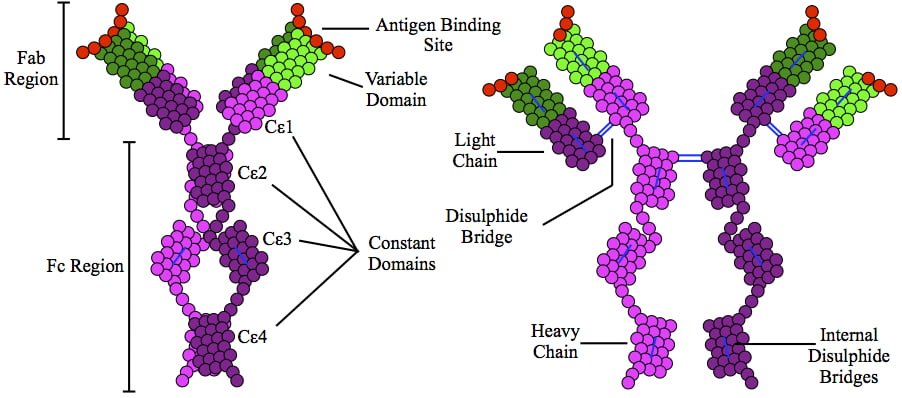
This is a depiction of immunoglobulin E (IgE) antibody which is elevated in allergic reactions.
What Is Allergic Rhinitis?
A llergic rhinitis is an immune disorder characterized by inflammation of the nasal mucosa by an IgE antibody reaction to an allergen.
An allergen is something that triggers an allergic immune response.
Q: What is the immune response?
A: Implicated in the response is an increase in T gamma-delta intraepithelial lymphocytes (IELs), which is a subset of pro-inflammatory T-cells located in the respiratory mucosa. Lymphocytes are white blood cells.
When a person with allergic rhinitis breathes in an allergen such as pollen or dust, the body releases chemicals, including histamine that cause allergy symptoms. For example, hay fever involves an allergic reaction to pollen. A similar reaction occurs with allergy to mold, animal dander, dust, and other allergens that are breathed in.
What Is Allergic Rhinitis In Celiac Disease and/or Gluten Sensitivity?
Drawing shows changes in airways during asthma attack. wikipedia
What Is Asthma?
Asthma is a chronic immune respiratory condition characterized by narrowing and inflammation of the lung airways (large bronchi, bronchial tubes and small bronchioles) in response to an allergen as the trigger or stimulus. As such, asthma occurs in episodes and does not result in progressive loss of pulmonary function.
During an asthma attack, airways constrict, trapping air so lungs become overinflated. Normally, bronchial airways bring air to millions of air sacs that are attached to the ends of bronchioles. Air sacs, called alveoli, are only one cell thick to allow for rapid exchange of gases.
That is, oxygen from air breathed into the sacs moves into the bloodsteam and carbon dioxide is released from the bloodstream to air that is breathed out of air sacs.
The outer walls of bronchioles are made up of muscles which, in the process of breathing, normally contract on expiration to help expel air and then relax. During an asthma attack, these muscles abnormally constrict, impairing airflow into and out of the alveoli. This is called bronchospasm.
Common allergens that cause inflammation include airborne dust mite feces, mold, and pollen and foods such as wheat, cow’s milk, eggs, and peanuts. Non-allergenic triggers include exercise, air pollution, smoking, and viral respiratory infection.
Q: What effect does inflammation have on the lungs?
A: Inflammation causes local tissue edema or swelling of the bronchioles and mucus formation. Inflammation with increased mucus secretions and edema narrows the airways that connect to alveoli which makes breathing difficult. Two things happen:
Inflammation impairs exchange of gases in alveoli, resulting in lack of sufficient oxygen (O2) for body cell functions, called hypoxia, and build-up of carbon dioxide (CO2) in blood, called CO2 retention.
Inflammation narrows passageways because of swelling, which reduces the movement of air to and from the alveoli through the airways, and this puts stress on the right side of the heart.
Treatment is aimed at controlling bronchospasm and reducing inflammation. Untreated asthma can be disabling and life threatening.
What Is Asthma In Celiac Disease and/or Gluten Sensitivity?
Prolactin Structure. Courtesy Boris TM at Wikipedia.
What Is Hyperprolactinemia?
H yperprolactinemia is an excess secretion of prolactin hormone causing an elevated blood level that is characterized by its effects on reproduction: in females causing amenorrhea and in males causing reduced sexual potency.
Q: What is prolactin?
A: Prolactin is a hormone produced by the the anterior pituitary gland that is transported by the bloodstream to stimulate androgen hormone production in male and estrogen hormone production in female sex glands. However, too much prolactin inhibits hormone production of estrogen in women and testosterone in men.
In pregnant females upon delivery of a baby, prolactin stimulates breast development and milk production for the purpose of feeding the infant. Sucking by the infant stimulates the release of prolactin which keeps the milk supply adequate.
Prolactin also promotes autoimmune mechanisms. Prolactin interferes specifically with B cell tolerance induction, enhances proliferative response to antigens and mitogens and increases the production of immune globulins, cytokines and autoantibodies.1
Other conditions that can stimulate prolactin release include nutritional deficiencies, thyroid stimulating hormone (TSH) produced by the pituitary gland, stress, exercise, and anterior pituitary tumors.
What Is An Elevated Prolactin Level In Celiac Disease and/or Gluten Sensitivity?
Sources:
Shelly S, Boaz M, Orbach H. Prolactin and autoimmunity. Autoimmun Rev. 2012 May;11(6-7):A465-70. doi: 10.1016/j.autrev.2011.11.009. [↩]
A feature of osteomalacia characterized by painful proximal muscle weakness involving the thighs and upper arms and hyperreflexia resulting from deficiencies of phosphorus and vitamin D.

 Osteomalacia is common in celiac disease. Osteomalacia can occur at any age. It children, it is called rickets. It is a metabolic bone disorder that involves slow loss of minerals from bone tissue throughout the skeleton, stemming from inadequate absorption of vitamin D. As minerals are dissolved from bone tissue to provide for essential functions elsewhere in the body, bones gradually lose their hardness.