Skip to content

Magnesium Deficiency

magnesium
Bananas Are a Good Source of Natural Magnesium.

What Is Magnesium?

[dropcap]M[/dropcap]agnesium is an essential mineral predominantly found in the body within cells, where it is vital for their functions.

Here is a summary of what magnesium does in our body:

  1. Co-factor for over 300 enzymes involved in the metabolism of food components and synthesis of many compounds.
  2. Required for nerve transmission.
  3. Required for muscle activity (acts to relax muscles in opposition to calcium which acts to contract).
  4. Acts to maintain heart rhythm.
  5. Required for membrane transport and interactions.
  6. Required for glucose metabolism and energy production within cells.
  7. Component of bone mineralization and tooth formation.
  8. Plays a key role in calcium and phosphorus metabolism and management by parathyroid hormone.
  9. Maintains the structural and functional integrity of vital eye tissues such as lens.1

Only 1% of magnesium is found in blood, but the body must keep blood levels of magnesium constant.

What Is Magnesium Deficiency In Celiac Disease and/or Gluten Sensitivity?

Sources:
  1. Agarwal R, Iezhitsa I, Agarwal P, Spasov A. Magnesium deficiency: does it have a role to play in cataractogenesis? Exp Eye Res. 2012 Aug; 101:82-9. doi: 10.1016/j.exer.2012.05.008. []

Chronic Fatigue Syndrome 

What Is Chronic Syndrome Fatigue? [dropcap]C[/dropcap]hronic fatigue syndrome is a debilitating illness characterized by persistent or relapsing overwhelming and incapacitating fatigue not relieved by rest, having a definite onset and often accompanied by numerous symptoms… 

Vitamin D Deficiency

Sunshine Delivers Free Vitamin D To Everyone.

What Is Vitamin D?

[dropcap]V[/dropcap]itamin D is the principle regulator of calcium homeostasis (balance) in the body.  This “vitamin” is really a prohormone, meaning it acts like a hormone but is not. Vitamin D does, however, contain cholesterol in its molecular structure like steroid hormones.

The physiological importance of vitamin D encompasses much more than the regulation of bone metabolism although this is a mighty function.

Q: How does vitamin D regulate bone metabolism?

A: In regulation of bone metabolism, vitamin D works in three ways: 1) enables active absorption of calcium from the small intestine, 2) enhances reabsortion of calcium by the kidneys that would otherwise be excreted in urine, and 3) plays an active role in skeletal development and bone mineralization. Mineralization gives strength to living bone tissue.

Vitamin D interacts with receptors within cells to effect transcriptional changes in many cell types including those in gut, bone, breast, prostate, brain, skeletal muscle, and the immune system.1

In regards to the essential role of vitamin D in muscle tissue, it has been recently shown that vitamin D regulates both muscle function and structure of primary myofibers.2

Vitamin D is converted in the body to a molecule that is biologically active. The active form is 1,25-dihydroxyvitamin D, usually referred to as vitamin D3. About 80% comes from sun exposure and the remaining from food.

Vitamin D3 is synthesized in the skin from 7-dehydrocholesterol via photochemical reactions requiring UV light (sunlight). That is, light that contains energy from the sun is incorporated into molecules of  7-dehydrocholesterol in the underlying dermis of skin to make this vitamin. This is why inadequate exposure to sunlight contributes to vitamin D deficiency.

Blood concentration of 25(OH)D is the best indicator of vitamin D status. It reflects vitamin D produced in the skin and that obtained from food and supplements and has a fairly long circulating half-life of 15 days.3

What Is Vitamin D Deficiency In Celiac Disease and/or Gluten Sensitivity?

Sources:
  1. McCarty DE, Chesson AL Jr, Jain SK, Marino AA. The link between vitamin D metabolism and sleep medicine. Sleep Med Rev. 2014 Aug;18(4):311-9. doi: 10.1016/j.smrv.2013.07.001. []
  2. Girgis CM, Mokbel N, Cha KM, Houweling PJ, Abboud M, Fraser DR, Mason RS, Clifton-Bligh RJ, Gunton JE. The vitamin D receptor (VDR) is expressed in skeletal muscle of male mice and modulates 25-hydroxyvitamin D (25OHD) uptake in myofibers. Endocrinology. 2014 Sep;155(9):3227-37 []
  3. http://ods.od.nih.gov/factsheets/VitaminD-HealthProfessional/ []

Hypomagnesemia (Low Blood Level of Magnesium)

hypomagnesemiaWhat Is Hypomagnesemia?

[dropcap]H[/dropcap]ypomagnesemia means the level of magnesium in the bloodstream is too low to meet metabolic needs of the body for this mineral.

Q: What are the metabolic needs of the body for magnesium?

A: The metabolic needs of the body for magnesium are numerous which gives rise to very many distressing symptoms when this mineral is deficient.

A major function of magnesium is to stabilize the structure of an enzyme called adenosine triphosphate (ATP) within cells for the production of energy. In the brain, magnesium plays important roles in all the major metabolisms such as oxidation-reduction and regulation of ions (charged minerals).1

What Is Hypomagnesemia In Celiac Disease and/or Gluten Sensitivity?

Sources:
  1. Bourre JM. Effects of nutrients (in food) on the structure and function of the nervous system: update on dietary requirements for brain. Part 1: micronutrients. J Nutr Health Aging. 2006 Sep-Oct;10(5):377-85. []

Dermatomyositis

dermatomyositisWhat Is Dermatomyositis?

[dropcap]D[/dropcap]ermatomyositis is a rare autoimmune systemic disease of the connective tissue that is characterized by inflammatory and debilitating degenerative changes in the muscles and in the skin. 

Dermatomyositis results in symmetric, proximal muscle weakness of limbs (upper arms and legs), and skin manifestations. 50-70% of patients have circulating myositis-specific auto-antibodies.

The course of dermatomyositis is unpredictable being marked by spontaneous flare-ups and remissions. It can begin slowly or abruptly according to the factor that is triggering the onset such as infection, medications like phenytoin, and autoimmune disease.

Q: What are the skin manifestations of dermatomyositis?

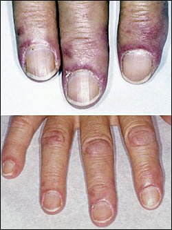
A: Classic skin manifestations of dermatomyositis include these features:

  • The heliotrope rash (lilac color) on upper eyelids.
  • Rash on face, neck, shoulders, upper chest, elbows, knees, knuckles, and back.
  • Gottron’s papules (scaly, red eruptions or  patches over the knuckles, elbows, and knees).
  • The V-sign (rash front of neck and chest).
  • The shawl sign (rash distribution on shoulders and back).1

Additional cutaneous manifestations are described below under symptoms.

Dermatomyositis is associated with an increased risk of  cancer, other autoimmune diseases, such as lupus and psoriasis, and it can be a complication of interferon-α therapy. About 1 person in 100,000 are affected according to various studies. While it affects all ages, women have twice the occurence of men.

There is no cure for dermatomyositis, but the symptoms can be treated.  Options include medication, physical therapy, exercise, heat therapy (including microwave and ultrasound), orthotics and assistive devices, and rest.  The standard treatment for dermatomyositis is a corticosteroid drug, given either in pill form or intravenously.  Immunosuppressant drugs, such as azathioprine and methotrexate, may reduce inflammation in people who do not respond well to prednisone.2

What Is Dermatomyositis In Celiac Disease and/or Gluten Sensitivity?

Sources:
  1. Marvi U, Chung L, Fiorentino DF. Clinical presentation and evaluation of dermatomyositis. Indian J Dermatol. 2012 Sep;57(5):375-81. doi: 10.4103/0019-5154.100486. []
  2. National Institute of Neurological Disorders and Stroke. []

Hashimoto’s Disease (Autoimmune Thyroiditis Causing Hypothyroidism)

DyspareuniaWhat Is Autoimmune Thyroiditis (Hypothyroidism)?

[dropcap]A[/dropcap]utoimmune thyroiditis, also called Hashimoto’s thyroiditis or Hashimoto’s Disease, is an autoimmune destruction of thyroid tissue characterized by insufficient thyroid hormone circulating in the body that causes formation of a goiter (enlarged thyroid gland) and hypothyroidism.

Hypothroidism refers to the condition of markedly reduced secretion of thyroid hormone. There are other causes of hypothyroidism besides Hashimoto’s thyroiditis.

Hashimoto’s thyroiditis is often associated with other autoimmune diseases such as celiac disease.

In Hashimoto’s thyroiditis, a profusion of antibodies are produced, which build up in the blood. Left untreated so that much of the thyroid gland is destroyed, this condition may progress to the very serious and life-threatening condition called myxedema.

Note: In myxedema, protein, electrolytes, and water abnormally accumulate in between cells which produce firm, inelastic puffy skin that is cool, dry, rough, scaly, and may appear yellow; in some people, areas such as the ankles become crusty with a look of tree bark. Many systemic changes develop shown by significant slowing of mental and physical functions. Please see below.

Q: What thyroid tissue is targeted for destruction?

A: In Hashimoto’s thyroiditis, high levels of autoantibodies target thyroglobulin and thyroid peroxidase, leading to inflammation and destruction of the thyroid gland. The resulting fibrosis or scarring of the gland results in lack of thyroid hormone production.

The thyroid gland consists of a large number of closed vesicles that contain a homogenous substance called colloid, which contains the thyroglobulin. Thyroglobulin is an iodine-containing protein secreted by the thyroid gland and stored within its colloid, from which the thyroid hormones thyroxine (T4) and triiodothyroinine (T3) are derived.1

T3 is the active hormone and is made from T4. Thyroid hormones affect metabolism, brain development, breathing, heart and nervous system functions, body temperature, muscle strength, skin dryness, menstrual cycles, weight, and cholesterol levels.

Thyroid hormone production is regulated by thyroid-stimulating hormone (TSH), which is made by the pituitary gland in the brain. Normally, when thyroid hormone levels in the blood are low, the pituitary releases more TSH. When thyroid hormone levels are high, the pituitary decreases TSH production.

Hashimoto’s disease, with or without the development of hypothyroidism, is treated with synthetic thyroxine, which is man-made T4. Health care providers prefer to use synthetic T4, such as Synthroid® (Levothyroxine), rather than synthetic T3, because T4 stays in the body longer, ensuring a steady supply of thyroid hormone throughout the day. The thyroid preparations made with animal thyroid are not considered as consistent as synthetic thyroid.2

What Is Autoimmune Thyroiditis In Celiac Disease and/or Gluten Sensitivity?

Sources:
  1. Taber’s Cyclopedic Medical Dictionary. 19th ed. F.A. Davis Company. Philadelphia, PA. []
  2. National Endocrine and Metabolic Diseases Information Service. []

Polymyositis

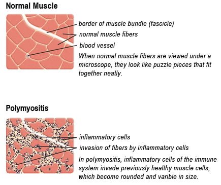
Drawing of Biopsy Showing Muscle Fibers Invaded by Immune Cells. Courtesy MDA.org

What Is Polymyositis?

[dropcap]P[/dropcap]olymyositis is a body-wide connective tissue disease resulting from autoimmune attack of skeletal muscles that is characterized by inflammatory and degeneratory changes. The course is unpredictable being marked by spontaneous flare-ups and remissions.

Polymyositis can begin slowly or abruptly according to the factor that is triggering the onset such as infection, medications like phenytoin, and autoimmune disease.

Progressive muscle weakness starts in the proximal skeletal muscles (muscles closest to the trunk of the body).

Skeletal muscles, also called voluntary, are muscles that move the body as we want, such as walking and  lifting objects, as opposed to those we cannot voluntarily control, such as the muscles of digestion. 

Q: What are the degeneratory changes in skeletal muscles?

A: In polymyositis, degeneratory changes in skeletal muscles means that muscles are being destroyed (called necrosis), resulting in fibrosis, or scarring. When scar tissue takes the place of lost muscle tissue, it cannot act like muscle to contract and relax.   Muscle destruction is what causes muscle pain and weakness.

After the clinical work-up of exams and blood studies to determine muscle damage, the diagnosis of polymyositis is confirmed by muscle biopsy. See image at above left. The black dots are inflammatory cells. Edema (fluid) between cells caused by inflammation pushes muscle fibers apart.

There is no cure for polymyositis, but the symptoms can be treated.  Options include medication, physical therapy, exercise, heat therapy (including microwave and ultrasound), orthotics and assistive devices, and rest.  The standard treatment for polymyositis is a corticosteroid drug, given either in pill form or intravenously.  Immunosuppressant drugs, such as azathioprine and methotrexate, may reduce inflammation in people who do not respond well to prednisone. 

Periodic treatment using intravenous immunoglobulin can also improve recovery.  Other immunosuppressive agents used to treat the inflammation associated with polymyositis include cyclosporine A, cyclophosphamide, and tacrolimus.  Physical therapy is usually recommended to prevent muscle atrophy and to regain muscle strength and range of motion.1

Diagnosis is based on elevated muscles enzymes, increased urinary creatine level, and electromyograph abnormalities.

Polymyositis can affect people at any age. It is most common in adults between ages 50 and 70, and in children ages 5 to 15. It affects women twice as often as men and is more common in African Americans than Caucasians.2 The major causes of death from polymyositis are cancer and lung disease, including pneumonia.  The 5-year mortality rate can be as high as 1 in 5 patients.2

What Is Polymyositis In Celiac Disease and/or Gluten Sensitivity?

Sources:
  1. National Institute of Neurological Disorders and Stroke []
  2. http://www.nlm.nih.gov/medlineplus/ency/article/000428.htm [] []

Tetany

Trouseau Testing For Tetany.
Trouseau Testing For Tetany. Courtesy Digplanet.

What Is Tetany?

[dropcap]T[/dropcap]etany is a painful feature of low blood calcium or low blood magnesium causing hyperexcitability of all nerves which in turn stimulates involuntary sudden, intermittent and repetitious tonic spasms of muscles. Tonic spasms are steady rather than jerky.

Causes include low function of the parathyroid glands which regulate calcium blood levels, vitamin D deficiency which inhibits calcium absorption from the gut that leads to calcium deficiency, and alkalosis.

In alkalosis, the chemical pH (acid/alkaline measure) of blood is too high, or alkaline, which inhibits adequate free calcium ions from being available for cell use.

Q: Why do low blood levels of calcium or magnesium cause muscles to spasm?

A: Calcium controls the readiness of muscle to respond to nerve stimulation. In muscle action, calcium and magnesium are minerals that oppose each other. Calcium allows a muscle to contract while magnesium allows it to relax. Inadequate levels of these opposing minerals affect the ability of muscles to properly function.

A simple test for tetany is called Trouseau’s named after the doctor who developed it. A blood pressure cuff is placed on an upper arm as usual and pumped up to a pressure sufficient to stop blood flow.  The pressure must be held for 3 minutes. A positive result is shown in the above photo of carpopedal spasm: the wrist and hand flexes inward while the fingers extend (opposite of wrist and hand) and the thumb goes against the palm.

What Is Tetany In Celiac Disease and/or Gluten Sensitivity?

Selenium Deficiency

selenium gluten free celiac disease symptomWhat Is Selenium?

[dropcap]S[/dropcap]elenium is a mineral that is required by the body  in trace amounts for a healthy immune system, normal thyroid function, and antioxidant protection.

Selenium is absolutely required in the production of at least 30 selenoproteins in the body. First, selenium is joined to the amino acids cysteine as selenocysteine and to methionine as selenomethionine before being used as components for selenoproteins. Many selenoproteins are important antioxidant enzymes such as glutathione peroxidase.

Q: How does glutathione peroxidase work?

A: Glutathione peroxidase activity helps the recycling of vitamins C and E in optimizing the performance of the antioxidant system. The antioxidant properties of selenoproteins help prevent cellular damage from free radicals. Free radicals are natural by-products of oxygen metabolism that may contribute to the development of chronic diseases such as cancer and heart disease.

In the immune system, selenium stimulates immune properties of lymphocytes (white blood cells) by contributing to higher natural killer lymphocyte activity. Natural killer lymphocytes have the ability to destroy cancer cells and bacterial and viral agents.

Other selenoproteins help protect the thyroid gland from anti-oxidants and regulate thyroid function. Specifically, selenium plays an integral role in thyroid gland metabolism.1 Functions are more fully described below.

According to the Food and Agriculture Organization, United Nations, approximately 30 percent of tissue selenium is contained in the liver, 15 percent in kidney, 30 percent in muscle, and 10 percent in blood plasma.

What Is Selenium Deficiency In Celiac Disease and/or Gluten Sensitivity?

Sources:
  1. Stazi AV, Trinti B. Selenium status and over-expression of interleukin-15 in celiac disease and autoimmune thyroid diseases. Ann Ist Super Sanita. 2010;46(4):389-99.DOI: 10.4415/ANN_10_04_06. []