Skip to content

Ichthyosis, Acquired  

cheilosisWhat Is Cheilosis?

C heilosis is a painful feature of nutritional deficiencies, especially riboflavin and/or pyridoxine deficiency, and less frequently, from vitamin B12, folic acid, and iron. It is characterized by redness of the lips with cracking and weeping in the corner of the mouth.

Q: What is the consequence of weeping at the corners of the mouth?

A: The open sores that develop at the corners of the mouth invite infection by candida albicans, a yeast organism, and less commonly by bacteria normally present on the skin such as staphylococcus aureus.

These sores make opening the mouth very painful and so restrict eating, talking, socializing, and dental care.

What Is Cheilosis In Celiac Disease and/or Gluten Sensitivity?

Melanoma

Enamel_celiac[1]What Are Dental Enamel Defects?

D ental enamel defects (DED) comprise a variety of abnormalities in the dental enamel of the second or permanent teeth and of primary or baby teeth.

Teeth may appear with pits, thin or missing enamel, non-white enamel (yellow, orange, or gray colored teeth), and demarcated opacities (very white spots) even if teeth are white.

Q: What is enamel?

A: Dental enamel is the shiny, hard, white, dense, inorganic substance covering the crowns of the teeth. The crown is the tooth portion above the gum. Under the enamel is hard dentin which surround the living pulp. In teeth with missing enamel, the areas with exposed dentin appear dull and tan colored.

What Are Dental Enamel Defects In Celiac Disease and/or Gluten Sensitivity?

Pityriasis Rubra Pilaris

Vitamin C Deficiency: Clean Teeth with Swollen Gums.
Vitamin C Deficiency: Swollen Gums.

What Are Bleeding / Swollen Gums?

B leeding and/or swollen gums is  an inflammatory disorder characterized by changes in gum integrity that may result in tooth loss.

Sponginess of the gum (swelling) indicates inflammation, called gingivitis. 

Q: What causes gingivitis?

A: Gingivitis that is caused by inadequate dental hygiene shows as pockets of swollen gum between or along the base of teeth where sticky plaque was allowed to build up. Plaque holds bacteria that inflames the gums where it occurs.

Gingivitis that is caused by vitamin C deficiency, pregnancy, certain drugs such as the anti-epileptic drug phenytoin, and leukemia affects the whole gum irrespective of mouth care.

What Are Bleeding / Swollen Gums In Celiac Disease and/or Gluten Sensitivity?

Itchy (Pruritic) Skin  

tongue in B12 deficiencyWhat Is Beefy Red, Smooth, Burning Tongue?

B eefy, red, smooth, burning tongue is an inflammatory alteration in tongue tissue characteristic of vitamin B12 deficiency.

Inflammation makes the tongue red and sore which, when untreated, progresses to atrophied papillae (shrunken taste buds) that makes the tongue smooth.

Q: Why does vitamin B12 alter the tongue?

A: Vitamin B12 is essential for the metabolism of all cells, especially for those of the digestive tract of which the tongue is an organ.1

These easily seen changes to tongue tissue are an important diagnostic feature of deficient vitamin B12, which causes other devastating unseen damage, because the discomfort generally causes the individual to seek medical care.

What Is Beefy Red, Smooth, Burning Tongue In Celiac Disease and/or Gluten Sensitivity?

Sources:
  1. Krause’s Food, Nutrition, & Diet Therapy. 10th Edition. Kathleen Mahan, Sylvia Escott-Stump. 2000. W.B. Saunders Company. []

Lymphoma, B-Cell Non-Hodgkin’s

e3343bf0a689b5f974a852fa2e84a718What Is Laryngospasm?

L aryngospasm is an acute disorder of the larynx that is characterized by life-threatening spasms of the muscles of the larynx located in the throat.

Laryngospasm is a rare feature of hypocalcemia (low blood calcium level) characterized by severe alteration in nerve conduction and muscle contraction.

What Is Laryngospasm In Celiac Disease and/or Gluten Sensitivity?

Diabetic Instability 

Vertebrae. Courtesy FreePik.com

What Is Elevated Bone Alkaline Phosphatase?

E levated bone alkaline phosphatase (BALP) is a laboratory result that indicates an abnormal blood level of this bone enzyme.

A bone alkaline phosphatase blood level is one of the most frequently used biochemical markers of bone formation.

Q: Why is the purpose of  bone alkaline phosphatase?

A: Bone alkaline phosphatase is produced by bone cells called osteoclasts in normal bone maintenance for the purpose of breaking down old or damaged bone so that other bone cells called osteoblasts can fill in the excavated areas with new bone. This process keeps bone stong and healthy.

Elevated bone alkaline phosphatase shows that more bone is being broken down than is being replaced. It can be caused by hyperparathyroidism, bone tumors from cancer, and malnutrition.

What Is Elevated Bone Alkaline Phosphatase (BALP) In Celiac Disease and/or Gluten Sensitivity?

Hypokalemic Rhabdomyolysis in Celiac Disease and Dermatitis Herpetiformis

mucosal web in plummer-vinson syndromeWhat Is Plummer-Vinson Syndrome Affecting the Esophagus?

P lummer-Vinson syndrome is a manifestation of severe, long-term, iron deficiency anemia that is characterized by post-cricoid esophageal webs and dysphagia.

Q: What are esophageal webs?

A: Esophageal webs are one or more thin horizontal membranes consisting of squamous epithelium (cells that line the surface of the esophagus) and submucosa. They usually protrude from the anterior (front) wall, extending laterally across the inside esophagus but not to the posterior (rear) wall, which means that they rarely encircle the lumen.1

Dysphagia, or difficulty swallowing, from these webs is commonly painless and intermittent or progressive and may cause obstruction.

Webs can be detected by barium swallow X-ray, but the best way for demonstration is videofluoroscopy and by upper gastrointestinal endoscopy. They appear smooth, thin, and gray with eccentric or central lumen space. The webs typically occur in the upper part of the esophagus and may be missed and accidentally ruptured unless the endoscope is introduced under direct visualization.1

Iron deficiency is believed to decrease the contraction amplitude or force of the esophageal muscle resulting in motility impairment. Slower transit times have been recorded at the proximal and middle parts of the esophagus of Plummer-Vinson syndrome patients compared to healthy volunteers.2 Transit time is how fast ingested food and fluids travel through the esophagus.

Gude et al, report that iron replacement does not necessarily reverse the dysphagia in all the cases of Plummer-Vinson syndrome and that close monitoring of the web is mandated to watch for malignant change.  In fact, 3 to 15 per cent of the patients with Plummer-Vinson syndrome, mostly women between 15 and 50 years of age, have been reported to develop esophageal or pharyngeal cancer.2

What Is Plummer-Vinson Syndrome Affecting the Esophagus In Celiac Disease and/or Gluten Sensitivity?

Sources:
  1. Novacek G. Plummer-Vinson syndrome. Orphanet J Rare Dis. 2006; 1: 36. Published online 2006 September 15. doi:  10.1186/1750-1172-1-36. [] []
  2. Gude D, Bansal DP, and Malu A. Revisiting Plummer Vinson Syndrome. Annals of Medical and Health Sciences Research. 2013 Jan-Mar;3(1):119-121. [] []

Primary Hyperparathyroidism

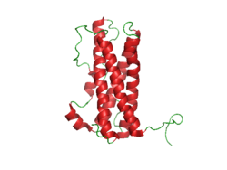
Prolactin Structure. Courtesy Boris TM at Wikipedia.

What Is Hyperprolactinemia?

H yperprolactinemia is an excess secretion of prolactin hormone causing an elevated blood level that is characterized by its effects on reproduction: in females causing  amenorrhea and in males causing reduced sexual potency.

Q: What is prolactin?

A: Prolactin is a hormone produced by the the anterior pituitary gland that is transported by the bloodstream to stimulate androgen hormone production in male and estrogen hormone production in female sex glands. However, too much prolactin inhibits hormone production of estrogen in women and testosterone in men.

In pregnant females upon delivery of a baby, prolactin stimulates breast development and milk production for the purpose of feeding the infant. Sucking by the infant stimulates the release of prolactin which keeps the milk supply adequate.

Prolactin also promotes autoimmune mechanisms. Prolactin interferes specifically with B cell tolerance induction, enhances proliferative response to antigens and mitogens and increases the production of immune globulins, cytokines and autoantibodies.1

Other conditions that can stimulate prolactin release include nutritional deficiencies, thyroid stimulating hormone (TSH) produced by the pituitary gland, stress, exercise, and anterior pituitary tumors.

What Is An Elevated Prolactin Level In Celiac Disease and/or Gluten Sensitivity?

Sources:
  1. Shelly S, Boaz M, Orbach H. Prolactin and autoimmunity. Autoimmun Rev. 2012 May;11(6-7):A465-70. doi: 10.1016/j.autrev.2011.11.009. []

Hypoparathyroidism, Idiopathic

hypocalcemia celiac disease gluten symptomWhat Is Hypocalcemia?

H ypocalcemia, or low plasma calcium, means the level of calcium in blood is too low to meet metabolic needs of the body for calcium.

Low blood calcium is characterized by bone and tooth demineralization (loss of calcium causing weak teeth and fragile bones), and these impaired functions: nerve conduction, muscle contraction, blood clotting, blood pressure regulation, glycogen to glucose conversion, many hormone actions, many enzyme activities, and acetylcholine production.

Q: Where is calcium found in the body?

A: Calcium is the most abundant mineral in the body, with 99% residing in bones and teeth where it constitutes 40% of skeletal bone weight along with 45% phosphorus. As a component of bone (hard tissue), calcium fulfills a structural role to maintain body size and act as attachments for musculoskeletal tissues. The remaining 1% of calcium is present in blood and soft tissues.

Calcium levels in the blood are maintained within very strict limits by dietary intake, hormonal regulation by the parathyroid gland and a rapidly exchangeable pool in bone tissue.

What Is Hypocalcemia In Celiac Disease and/or Gluten Sensitivity?

Nails, Rounded With Curved Ends

What Is Angina Pectoris?Coronary Artery Lesion

A ngina pectoris, or simply angina, is a coronary syndrome characterized by an oppressive substernal pain (pain under breastbone) or pressure brought on by exertion and relieved by rest that results from failure of coronary arteries to deliver adequate oxygen to heart tissue due to ischemic heart disease.

Q: Why do coronary arteries fail to deliver adequate oxygen to heart tissue?

A: Coronary arteries are the blood vessels that serve the heart. In angina, these vessels fail to deliver adequate oxygen to heart tissue because they are narrowed or blocked by fatty buildups, called atherosclerotic plaques or by a blood clot which impair their ability to carry adequate blood that carries the oxygen. Diseased coronary arteries cannot deliver adequate oxygenated blood pumped by the heart to its own muscle cells.

The heart is a muscular organ that is working all the time without rest, so it needs a constant supply of oxygen. When heart muscle has to work harder, it needs more oxygen. Lack of oxygen causes pain which makes the affected person stop activity and rest.

Angina can be stable or unstable. Unstable angina is much more serious and can be life-threatening.

  • Stable angina produces predictable pain and responds to rest and/or medication. It is less serious than unstable angina but can be very painful or uncomfortable. Anything that makes the heart muscle need more oxygen can cause an angina attack in someone with heart disease, including: smoking, cold weather, exercise, emotional stress, obesity, and large meals. Other causes of angina include: abnormal heart rhythms (usually ones that cause the heart to beat quickly), anemia, coronary artery spasm, heart failure, heart valve disease, and hyperthyroidism (overactive thyroid).1
  • Unstable angina produces unpredictable pain that may occur at rest, lasting more than 20 minutes. It is more severe than stable angina and less responsive to medication. Atherosclerosis is by far the most common cause of unstable angina. Oxidized low-density lipoprotein, so-called bad cholesterol, and oxysterols play an important role in atherogenesis, the development of atherosclerosis. Coronary arteries that are narrowed by atherosclerotic plaques can rupture causing injury to the coronary blood vessel resulting in blood clotting which blocks the flow of blood to the heart muscle. Blood clots may form, partially dissolve, and later form again and angina can occur each time a clot blocks blood flow in an artery. People with unstable angina are at increased risk of having a heart attack.2

What Is Angina In Celiac Disease and/or Gluten Sensitivity?

Sources:
  1. http://www.ncbi.nlm.nih.gov/pubmedhealth/PMH0001247/ []
  2. http://www.heart.org/HEARTORG/Conditions/HeartAttack/SymptomsDiagnosisofHeartAttack/Unstable-Angina_UCM_437513_Article.jsp# []