Skip to content

Small Bowel Intussusception

Invagination_Schema[1]What Is Small Bowel Intussusception?

Small bowel intussusception is a bowel derangement that is characterized by the slipping of one section of intestine into another, leading to bowel obstruction.

Acute bowel intussusception is a rare manifestation in adults, which mainly involves the small intestine.1

What Is Small Bowel Intussusception In Celiac Disease and/or Gluten Sensitivity?

Sources:

  1. Grados A, Bernard F, Coquet-Reinier B, Rossi P, Bagneres D, Demoux AL, Marciano S, Frances Y, Granel B. Acute bowel intussusception revealing celiac disease a new case and literature review. Rev Med Interne. 2011 Oct;32(10):628-32. doi: 10.1016/j.revmed.2011.03.334 []
weight loss gluten free

Calories, Weight, and a Whole Lotta Garbage

I was looking back at old drafts of posts that I’ve written over the years but never published, and I found this rant on the 1200 calorie weight loss myth. Since it’s the last day of National Eating Disorders Awareness Week  and the first day of National Nutrition Month, I figured it would be a good time to share it. (Editor’s note: Originally published March 1, 2014)

weight loss gluten freeNot a week goes by that I don’t see a client who truly believes she/he needs to be following a 1200 calories diet to lose weight or be healthy. It seems to be the #1 nutrition myth.

Despite what the intertubes may say, a 1200 cal diet is not considered standard professional weight loss advice. Honest.There is, of course, plenty of bad advice and/or controversy out there, because that sells. There’s also a whole lot of standard nutrition advice that I heartily disagree with, but that’s another post. Take a  look at

Deal Alert! Gluten Free Medifast Meals: Free Shipping and Discounts for August

Free shipping and save up to $66 on Medifast’s gluten-free diet meal packages through August 31st.

Gluten-Free 4-Week Package

Medifast has developed two gluten-free packages for people on the gluten-free diet who want to lose weight quickly and safely. The gluten-free packages come in a 4 week package and a 2 week package.

Discount codes are good until August 31st, 2013.

Free shipping with a purchase of $150 or more: Click Here and use coupon code SHIPAUG13

Save $66 with a purchase of $250 or more: Click Here and use coupon code AUG31K

What is Medifast? From Medifast’s website…

Deal Alert! Gluten Free Medifast Meals: Free Shipping and Discounts for July

Free shipping and save up to $66 on Medifast’s gluten-free diet meal packages through July 31st.

Gluten-Free 4-Week Package

Medifast has developed two gluten-free packages for people on the gluten-free diet who want to lose weight quickly and safely. The gluten-free packages come in a 4 week package and a 2 week package.

Discount codes are good until July 31st, 2013.

Get Free Shipping with any purchase of $150 or more. Click here and use coupon code: SHIPJUL13

Save $66 with a purchase of $250 or more. Click here and use coupon code: JUL31K

What is Medifast? From Medifast’s website…

Dysgeusia (Impaired Taste)

DyspareuniaWhat Is Autoimmune Thyroiditis (Hypothyroidism)?

A utoimmune thyroiditis, also called Hashimoto’s thyroiditis or Hashimoto’s Disease, is an autoimmune destruction of thyroid tissue characterized by insufficient thyroid hormone circulating in the body that causes formation of a goiter (enlarged thyroid gland) and hypothyroidism.

Hypothroidism refers to the condition of markedly reduced secretion of thyroid hormone. There are other causes of hypothyroidism besides Hashimoto’s thyroiditis.

Hashimoto’s thyroiditis is often associated with other autoimmune diseases such as celiac disease.

In Hashimoto’s thyroiditis, a profusion of antibodies are produced, which build up in the blood. Left untreated so that much of the thyroid gland is destroyed, this condition may progress to the very serious and life-threatening condition called myxedema.

Note: In myxedema, protein, electrolytes, and water abnormally accumulate in between cells which produce firm, inelastic puffy skin that is cool, dry, rough, scaly, and may appear yellow; in some people, areas such as the ankles become crusty with a look of tree bark. Many systemic changes develop shown by significant slowing of mental and physical functions. Please see below.

Q: What thyroid tissue is targeted for destruction?

A: In Hashimoto’s thyroiditis, high levels of autoantibodies target thyroglobulin and thyroid peroxidase, leading to inflammation and destruction of the thyroid gland. The resulting fibrosis or scarring of the gland results in lack of thyroid hormone production.

The thyroid gland consists of a large number of closed vesicles that contain a homogenous substance called colloid, which contains the thyroglobulin. Thyroglobulin is an iodine-containing protein secreted by the thyroid gland and stored within its colloid, from which the thyroid hormones thyroxine (T4) and triiodothyroinine (T3) are derived.1

T3 is the active hormone and is made from T4. Thyroid hormones affect metabolism, brain development, breathing, heart and nervous system functions, body temperature, muscle strength, skin dryness, menstrual cycles, weight, and cholesterol levels.

Thyroid hormone production is regulated by thyroid-stimulating hormone (TSH), which is made by the pituitary gland in the brain. Normally, when thyroid hormone levels in the blood are low, the pituitary releases more TSH. When thyroid hormone levels are high, the pituitary decreases TSH production.

Hashimoto’s disease, with or without the development of hypothyroidism, is treated with synthetic thyroxine, which is man-made T4. Health care providers prefer to use synthetic T4, such as Synthroid® (Levothyroxine), rather than synthetic T3, because T4 stays in the body longer, ensuring a steady supply of thyroid hormone throughout the day. The thyroid preparations made with animal thyroid are not considered as consistent as synthetic thyroid.2

What Is Autoimmune Thyroiditis In Celiac Disease and/or Gluten Sensitivity?

Sources:
  1. Taber’s Cyclopedic Medical Dictionary. 19th ed. F.A. Davis Company. Philadelphia, PA. []
  2. National Endocrine and Metabolic Diseases Information Service. []

Primary Hyperparathyroidism

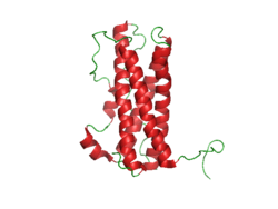
Prolactin Structure. Courtesy Boris TM at Wikipedia.

What Is Hyperprolactinemia?

H yperprolactinemia is an excess secretion of prolactin hormone causing an elevated blood level that is characterized by its effects on reproduction: in females causing  amenorrhea and in males causing reduced sexual potency.

Q: What is prolactin?

A: Prolactin is a hormone produced by the the anterior pituitary gland that is transported by the bloodstream to stimulate androgen hormone production in male and estrogen hormone production in female sex glands. However, too much prolactin inhibits hormone production of estrogen in women and testosterone in men.

In pregnant females upon delivery of a baby, prolactin stimulates breast development and milk production for the purpose of feeding the infant. Sucking by the infant stimulates the release of prolactin which keeps the milk supply adequate.

Prolactin also promotes autoimmune mechanisms. Prolactin interferes specifically with B cell tolerance induction, enhances proliferative response to antigens and mitogens and increases the production of immune globulins, cytokines and autoantibodies.1

Other conditions that can stimulate prolactin release include nutritional deficiencies, thyroid stimulating hormone (TSH) produced by the pituitary gland, stress, exercise, and anterior pituitary tumors.

What Is An Elevated Prolactin Level In Celiac Disease and/or Gluten Sensitivity?

Sources:
  1. Shelly S, Boaz M, Orbach H. Prolactin and autoimmunity. Autoimmun Rev. 2012 May;11(6-7):A465-70. doi: 10.1016/j.autrev.2011.11.009. []

Lymphoma, B-Cell Non-Hodgkin’s

e3343bf0a689b5f974a852fa2e84a718What Is Laryngospasm?

L aryngospasm is an acute disorder of the larynx that is characterized by life-threatening spasms of the muscles of the larynx located in the throat.

Laryngospasm is a rare feature of hypocalcemia (low blood calcium level) characterized by severe alteration in nerve conduction and muscle contraction.

What Is Laryngospasm In Celiac Disease and/or Gluten Sensitivity?

Celiac Disease, Refractory

Anatomy of Upper Airway and Throat. Courtesy Wikipedia.org

What Is Post-Cricoid Carcinoma?

P ost-cricoid carcinoma is a rare malignancy arising in the hypopharynx.

Q: What is the hypopharynx?

A: The hypopharynx is the lower portion of the pharynx that opens into the larynx. It is comprised of the postcricoid region, pyriform sinus, and posterior hypopharyngeal wall.

Tumors rarely appear in the posterior pharyngeal wall or postcricoid region without also involving the pyriform sinus. The lack of anatomic barriers between these sites and a propensity for these tumors to develop in the pyriform sinus and spread outward account for this phenomenon.

Overall prognosis for these tumors is poor. Because of the rich organic lymphatic and vascular networks, aggressive growth and early cervical metastases (compared with cancers at other head and neck sites) characterize these cancers.

A study investigating the clinical course of 21 patients with post-cricoid carcinoma found the 5-year cause-specific survival rate was 52%. All the patients who eventually died did so within 3 years of the treatment. Seven patients had distant metastases, representing a higher frequency as compared with that of patients with recurrence of the primary focus and cervical lymph node metastasis. All of these patients who had been treated by surgery died of the cancer.1

Unfortunately, hypopharyngeal cancers are discovered at a later stage than other head and neck cancers. The delay in diagnosis from symptom onset averages 10 months.2

What Is Post-Cricoid Carcinoma In Celiac Disease and/or Gluten Sensitivity?

Sources:
  1. Asakage T, Nakao K, Ebihara Y, Fujishiro Y, Watanabe K. A clinical study of post-cricoid carcinoma. Acta Otolaryngol Suppl. 2007 Dec;(559):118-22. doi: 10.1080/03655230701599354. []
  2. http://emedicine.medscape.com/article/848359-overview []

Lymphomas, Extraintestinal

What Is Dysphagia? D ysphagia is a functional upper digestive disorder characterized by difficulty in swallowing or food getting stuck and swallowing that takes longer than 10 seconds to complete. Q: What determines the symptoms?… 

Primary Sclerosing Cholangitis 

A 3D Image From Magnetic Resonace Cholangiography. NIHMS
A 3D Image From Magnetic Resonace Cholangiography showing biliary tree. NIHMS

What Is Primary Sclerosing Cholangitis?

Primary sclerosing cholangitis (PSC) is an uncommon, slowly progressive bile duct disease that results in stagnation or build-up of bile in the liver, called cholestasis.

Primary sclerosing cholangitis is characterized by sclerosis, or scarring inflammation in bile ducts both within the liver (intra-hepatic ducts), and outside the liver (extra-hepatic ducts), causing progressive narrowing and, eventually, obliteration of the bile ducts.

Primary sclerosing cholangitis comes under the umbrella term autoimune liver disease in which the end result is immune-mediated hepatocellular (liver cell) or hepatobiliary (bile duct) injury.1

Q: What happens when scarred bile ducts can no longer transport bile out of the liver?

A: Bile that cannot be removed from the liver by the biliary duct system backs up and damages the liver, causing cirrhosis.

Bile is continually made by the liver from phospholipids salt, cholesterol, and aging blood cells that it removes from circulation to be carried out of the liver. Bile also carries away waste products produced by normal metabolism and toxic substances that are removed by the liver for eventual elimination in stool. As such, bile must continually flow out of the liver to prevent build-up in the liver.

Bile is a greenish brown liquid made by the liver. Bile ducts carry it out of the liver to the gall bladder for storage until needed to aid in the digestion and absorption of fat from the small intestine. Bile emulsifies fat eaten in the diet so that the pancreatic enzyme called lypase can break it down into its fatty acid and glycerol components.

The liver is the largest organ within the body. It lies mostly in the upper part of the abdomen on the right side just under the diaphragm. About 70% of liver tissue is made up of cube shaped cells called hepatocytes that do the main work of the liver. Other cells (epithelial) form structure and are arranged in single layers around blood vessels, sinusoids, and bile ducts. 

Build-up of bile in the liver is the end result of the inflammatory process in primary sclerosing cholangitis, that by swelling and scarring of bile ducts impedes and eventually prevents bile flow out of the liver, leading to liver failure. There is no curative treatment available for primary sclerosing cholangitis, besides liver transplantation.2

The appearance of the intrahepatic and extrahepatic biliary ducts can be assessed by use of cholangiography, and magnetic resonance (MR) imaging is the best way to identify patients.  See image above.3

MR cholangiography offers a noninvasive method of obtaining images of the biliary system without the use of a contrast agent. There is no radiation exposure. Pulse sequences can be chosen to obtain bright bile or black bile cholangiograms. Image processing algorithms can be selected to obtain a three-dimensional representation of biliary anatomy and pathology, and those images can be rotated in any plane so that ductal anatomy and pathology can be seen to best advantage.4

There is no cure for primary sclerosing cholangitis but there are symptom treatments one of which is supplementation for low levels of vitamins A,D,E, and K. Liver transplant is the only effective option.

What Is Primary Sclerosing Cholangitis In Celiac Disease and/or Gluten Sensitivity?

Sources:
  1. Trivedi PJ, Adams DH. Mucosal immunity in liver autoimmunity: a comprehensive review. J Autoimmun. 2013 Oct;46:97-111. doi: 10.1016/j.jaut.2013.06.013. []
  2. Kummen M, Schrumpf E, Boberg KM. Liver abnormalities in bowel diseases. Best Pract Res Clin Gastroenterol. 2013 Aug;27(4):531-42. doi: 10.1016/j.bpg.2013.06.013. []
  3. Eaton JE, Talwalkar JA, Lazaridis KN, Gores GJ, Lindor KD. Pathogenesis of primary sclerosing cholangitis and advances in diagnosis and management. Gastroenterology. 2013 Sep;145(3):521-36. doi: 10.1053/j.gastro.2013.06.052. Epub 2013 Jul 1. []
  4. Meakem TJ 3rd, Schnall MD. Magnetic resonance cholangiography. Gastroenterol Clin North Am. 1995 Jun;24(2):221-38. []