Skip to content

Antibody: definition
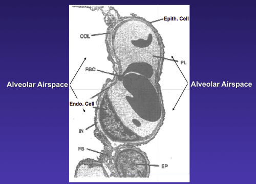
Image shows
Image Showing  Blood-Air Barrier in the Lung. Courtesy quizlet.com

What Is Increased Pulmonary Permeability?

[dropcap]I ncreased pulmonary permeability is a mucosal defect in the lung characterized by alteration of the normally tight epithelial  blood-air barrier in the lung apparently caused by inflammation.

Q: What is the blood-air barrier in the lung?

A: This blood-air barrier in the lung consists of the alveolar epithelium (surface cells of the alveoli), the underlying capillary endothelium (surface cells of the capillaries), their basement membranes and the interstitial space between the cell layers.

Alveoli are also called “air sacs.” The exchange of oxygen breathed into air sacs from air on inspiration and carbon dioxide breathed out from capillary blood on expiration occurs between  air sacs and capillaries.

The capillary endothelium prevents proteins in blood from leaking (permeating) into the air sacs while allowing water and small molecules to pass. This is why breath has moisture in it.

Little is known about the interactions between the alveolar and the blood compartment.1

What Is Increased Pulmonary Permeability In Celiac Disease and/or Gluten Sensitivity?

Sources:
  1. Neuhaus W, Samwer F, Kunzmann S, Muellenbach RM, Wirth M, Speer CP, Roewer N, Förster CY. Lung endothelial cells strengthen, but brain endothelial cells weaken barrier properties of a human alveolar epithelium cell culture model. Differentiation. 2012 Nov;84(4):294-304. doi: 10.1016/j.diff.2012.08.006. Epub 2012 Sep 27. []

Autoimmune Hepatitis: definition

Broken lower leg. Courtesy FreePik.com

What Are Bone Fractures?

[dropcap]B one fractures, or broken bones, are breaks in skeletal bones that occur usually from trauma to the bone itself or by a sudden violent contraction of muscle attached to it.

Q: Are there other causes of bone fractures besides trauma?

A: Fractures may occur spontaneously without trauma in certain pathological disorders such as osteoporosis, osteonecrosis, osteomalacia, osteomyelitis, syphilis, and cancer affecting the bone.

Bone fractures are a major public health problem with treatment costs in the billions of dollars and lead to subsequent disability for many patients.

Hip fractures, for example, may be complicated by infection at the surgical site, pneumonia, decubitus ulcer from lack of movement, and deep vein thrombosis making hip fractures the second leading cause of nursing home admissions in the USA.

Improvement of bone health and reducing risk factors such as smoking, caffeinated drinks (coffee, tea and sodas), and use of alcohol are key to preventing bone fractures.

Bone strength is easily measured by testing bone mineral density (BMD). BMD is evaluated by DEXA scan (dual-energy X-ray absorptiometry).  DEXA at the femoral neck and lumbar spine is considered the gold standard to confirm the diagnosis of osteoporosis.  Results are expressed as T and Z scores. T scores compare the result with a 20 to 40 year old helathy person while  Z scores compare the result with persons in the same age group. Both are measured in standard deviations (SD).

According to WHO criteria (World Health Organization), a T-score of -1 SD or greater denotes normal bone, a T-score between −1 to −2.5 SD denotes osteopenia, and a T-score of −2.5 or more denotes osteoporosis.1

What Are Bone Fractures In Celiac Disease and/or Gluten Sensitivity?

Sources:
  1. Pantaleoni S, Luchino M, Adriani A, Pellicano R, Stradella D, Ribaldone DG, Sapone N, Isaia GC, Di Stefano M, Astegiano M. Bone mineral density at diagnosis of celiac disease and after 1 year of gluten-free diet. ScientificWorldJournal. 2014;2014:173082. doi: 10.1155/2014/173082. []

Biopsy: definition

fimbria_ovaricaWhat Is Early Menopause?

[dropcap]E arly menopause is the permanent cessation of menstruation in females before the age of 40 years characterized by vasomotor instability (hot flashes/cold episodes), psychologic and emotional symptoms and profound changes in the lower genital tract.

The most severe forms present with absent pubertal development and primary amenorrhea termed primary ovarian failure (50% of these cases due to ovarian dysgenesis), whereas forms with post-pubertal onset are termed secondary ovarian failure and are characterized by disappearance of menstrual cycles associated with premature follicular depletion.1

Q: What is ovarian failure?

A: Ovarian failure is dysfunction of the ovaries to continue developing its eggs until about 51 years of age and to produce female hormones which results in infertility.  Ovarian failure  is  diagnosed by  this criteria:

  • High levels in the blood of follicle stimulating hormone secreted by the pituitary gland, and
  • Low levels of estrogen secreted by the ovaries.

The hypothalamus located in the brain is continually monitoring blood levels of estrogen so that when levels are low, it stimulates the nearby pituitary, or master gland, to secrete follicle stimulating hormone (FSH). FSH in turn stimulates the ovaries in females to produce estrogen. In ovarian failure, the ovaries do not respond to FSH. 

Some causes in the general population of women include cigarette smoking, viral infections, surgical removal of ovaries, chemotherapy, and autoimmune disorders such as celiac disease, Addison’s disease, hypoparathyroidism, diabetes mellitus, vitiligo, and pernicious anemia.

Who is Affected in the General Population? This heterogeneous disorder affects approximately 1% of women.2

What Is Early Menopause in Celiac Disease?

Sources:
  1. Beck-Peccoz P, Persani L. Premature ovarian failure. Orphanet J Rare Dis. 2006 Apr 6;1:9. []
  2. Beck-Peccoz P, Persani L. Premature ovarian failure. Orphanet J Rare Dis. 2006 Apr 6;1:9. []

Cholangiography: definition

intrauterine growth retardation gluten free
The twin on the right is much small than his brother on the left who has normal growth.

What Is Intrauterine Growth Retardation?

[dropcap]I ntrauterine growth retardation (IUGR) is a fetal development abnormality characterized by failure to grow normally for gestational period. Specifically, it means the developing baby weighs less than 90% of other babies at the same age.

Intrauterine growth retardation puts the baby at increased risk for complications such as premature birth or that the baby will die inside the womb before birth.1

Intrauterine growth restriction  may be suspected if the size of the pregnant woman’s uterus is small. The condition is usually confirmed by ultrasound. Further tests may be needed to screen for infection or genetic problems if intrauterine growth retardation is suspected.

Q: Why would a baby not grow normally during pregnancy? A: An unborn baby cannot grow normally  if it does not obtain adequate oxygen and nutrition delivered through the placenta from the mother. Factors that impede adequate delivery of nutrition include:

  • Poor placenta placement. Conditions that limit or interfere with space for nutrient and oxygen absorption between the placenta and the uterine wall include 1) low attachment of the placenta near or over the cervix where maternal blood supply is poor, 2) pulling away or bleeding between the placenta and uterine wall, 3) multiple placentas (from multiple babies) sharing the uterine wall may limit blood supply to one or more of the fetuses, and 4) the presence of an hydatid mole,  (non-fertilized egg growing wildly), tumor or fibroids taking up space or growing under or into the placenta. 
  • Chromosomal abnormalities in the fetus. Conditions such as trisomy 22  have early onset  of  intrauterine growth retardation  in pregnancy. 
  • Poor health of the mother. These factors include 1) anemia which impairs the ability of the mother’s blood to deliver adequate oxygen, 2) preeclampsia which interferes with placenta function, 3) diabetes which impairs proper supply of energy, 4) kidney disease, 5) poor diet, 6) malabsorption, 7) high blood pressure or heart disease, 8) clotting disorders, and 9) toxins and infections during pregnancy that may harm the developing baby such as rubella, cytomegalovirus, toxoplasmosis, and syphilis. 
  • Risk factors in the mother. Any of the following may contribute to intrauterine growth retardation:1
  • Alcohol abuse.
  • Drug addiction.
  • Smoking.

What Is Intrauterine Growth Retardation In Celiac Disease and/or Gluten Sensitivity?

Sources:
  1. http://www.nlm.nih.gov/medlineplus/ency/article/001500.htm [] []

Colic: definition

hypotoniaWhat Is Hypotonia?

[dropcap]H ypotonia is impairment  of muscle contraction that is characterized by abnormally low muscle tension or flaccid muscles. 

Normally, even when relaxed, muscles have a very small amount of contraction or tone that gives them a springy feel and provides some resistance to passive movement.  

Q: Why is muscle contraction impaired in hypotonia?

A: In hypotonia, muscle contraction is impaired due to a disconnect between nerve signals and responses of motor muscle. Motor muscles cause body parts to move such as the arms and legs.

Low muscle tone is not the same as low muscle strength, although these two conditions can co-exist.  Muscle tone is regulated by signals that travel from the brain to the nerves and stimulate the muscles to contract whereas muscle weakness is a problems of muscles themselves.

In babies this condition is called “floppy doll syndrome.”

What Is Hypotonia In Celiac Disease and/or Gluten Sensitivity?

Crohn: definition

A form of inflammatory bowel disease that causes irritation in the gastrointestinal (GI) tract. It usually affects the lower small intestine (also called the ileum) or the colon, but it can also affect any part…