Skip to content

Sjögren’s Syndrome 

Testing the Eyes for Sjogren's Syndrome.
Testing the Eye for Tear Production (L) and Damage to Conjunctiva from Dryness (R).

What Is Sjögren’s Syndrome?

[dropcap]S[/dropcap]jögren’s syndrome is a systemic inflammatory autoimmune disease with a chronic, progressive course that primarily attacks the lacrimal glands of the eye and the salivary glands of the mouth, which are exocrine glands. Exocrine glands secrete the substances they produce through a duct.

Sjögren’s syndrome is ordinarily characterized by dysfunction of the lacrimal glands to produce tears causing dry eye and the salivary glands to produce saliva causing dry mouth, but is not limited by or to these features.

Besides involvement of these exocrine glands, there may be involvement of other parts of the body, termed extraglandular, which may be more severe than eye or mouth features.

There is not yet agreement on classifying Sjögren’s syndrome. Primary and secondary are the two forms generally accepted.1 Both forms can cause mild to severe disease, called the spectrum:

  • Primary Sjögren syndrome. Disease occurs without involvement of other linked autoimmune disorders. In addition to the eyes and mouth, the nose, throat and skin may also be affected and joints, lungs, kidneys, blood vessels, digestive organs and nerves as well.2 Systemic manifestations (other than eyes and mouth) concern a third of patients, including lymphoma in 5% of the patients.3
  • Secondary Sjögren’s syndrome. Disease complicates other autoimmune disease such as systemic lupus erythematosus, rheumatoid arthritis, primary biliary cirrhosis, and celiac disease.

Diagnosis  of Sjögren’s syndrome is made by most doctors based on Schimer’s test for tears and unstimulated whole salivary flow to assess objective eye and oral involvement, since these are the tests most physicians use in clinical practice.4 Specific antibody tests would be  positive for anti-Ro (SSA)/anti-La (SSB) autoantibodies. Sjögren’s syndrome should also be considered when extraglandular manifestations such as vasculitis, polyneuropathy or arthritis occur, even when the patients do not complain of dry eyes and mouth.5

There is no cure for Sjögren’s syndrome. Treatment is aimed to diminish symptoms. For example, steroids and Ibupropen are used to decrease inflammation and pain in joints. Artificial tears and ointments are used for dry eye.

Most people who develop Sjogren’s syndrome are older than 40 years. Nine of ten people with Sjögren’s syndrome are women.2

What Is Sjögren’s Syndrome In Celiac Disease and/or Gluten Sensitivity?

Sources:
  1. Huang YF, Cheng Q, Jiang CM, An S, Xiao L, Gou YC, Yu WJ, Lei L, Chen QM, Wang Y, Wang J. The immune factors involved in the pathogenesis, diagnosis, and treatment of Sjogren’s syndrome. Clin Dev Immunol. 2013;2013:160491. doi: 10.1155/2013/160491. Epub 2013 Jul 9. []
  2. nlm.nih.gov [] []
  3. Fazaa A, Bourcier T, Chatelus E, Sordet C, Theulin A, Sibilia J, Gottenberg JE. Classification criteria and treatment modalities in primary Sjögren’s syndrome. Expert Rev Clin Immunol. 2014 Apr;10(4):543-51. doi: 10.1586/1744666X.2014.897230. []
  4. Cornec D, Saraux A, Cochener B, Pers JO, Jousse-Joulin S, Renaudineau Y, Marhadour T, Devauchelle-Pensec V. Level of agreement between 2002 American-European Consensus Group and 2012 American College of Rheumatology classification criteria for Sjogren’s syndrome and reasons for discrepancies. Arthritis Res Ther. 2014 Mar 19;16(2):R74. []
  5. Witte T. Pathogenesis and diagnosis of Sjögren’s syndrome. Z  Rheumatol. 2010 Feb;69(1):50-6. doi: 10.1007/s00393-009-0519-2. []

Candida Albicans Infection

Close-up shows inflammation and yellowish white patches of roof and back of the mouth caused by candidiasis. Uvula is greatly swollen, hanging near the level of the tongue. Courtesy: Wikipedia.
Close-up shows inflammation and infected patches on roof and back of the mouth caused by candidiasis. Uvula is swollen, hanging near the tongue. Courtesy: Wikimedia.

What Is Candida Albicans Infection?

[dropcap]C[/dropcap]andida albicans infection, called candidosis or candidiasis, is an opportunistic invasion of mucous membrane or skin by candida albicans, an endogenous yeast found in 40 to 80% of normal human beings. A former name for this small, budding fungus is monilia albicans.

Opportunistic means that yeast living on mucosal and skin surfaces does not invade (infect) unless these tissues become unhealthy and therefore cannot protect themselves.

Q: How does candida albicans cause infection?

A: Candida albicans lives on the mucosal surfaces and skin in most people without causing infection (colonizes) because of our normal defenses against invasion. In fact, candida albicans is a very effective colonizer of humans. For example, Russell and Lay found that 47% of 1-month-old infants were orally colonized with candida albicans, and 49% were colonized with other fungi.

During growth within the intestinal tract, the organism senses pH (acidity), oxygen, carbon sources, and the presence of surfaces allowing it to optimize gene expression for a particular environment. With these mechanisms for sensing, candida albicans is able to efficiently colonize humans in infancy.1

Candida Infection Of The Esophagus on X-ray. Courtesy Radiology Assistant.nl
Candida Infection Of The Esophagus (White Area) On X-ray. Courtesy Radiology Assistant.nl

Lowered host defenses allow yeast already present on mucosal and skin surfaces to take advantage and can grow rapidly, becoming pathogenic (disease producing) so that infection results.

Infection is characterized by superficial, irregular white patches on mucosal surfaces and possible invasion of the bloodstream by a filamentous form (thread-like structures) that can rapidly develop.

Candida albicans is unique among oral pathogens in its ability to invade cornified layers of stratified squamous epithelium of the tongue, mouth surfaces, hard and soft palate, esophagus, and gut. Stratified squamous epithelium is the tough surface cells that ordinarily protect underlying tissues from damage or invasion by microbes.

Candida albicans is also capable of invading the lungs and causing pneumonia and septicemia, which is the spread of infection into the bloodstream.

Here is a time honored simple do-it-youself test for infection of the mouth or throat: First thing in the morning before brushing your teeth or eating, fill a small see through glass with water then gently spit onto the surface. If after an hour the spit remains on the water surface, it is unlikely you have candida in the mouth. If it grows legs downward, it indicates that yeast is growing. If the spit sinks to the bottom, you have this problem. Yeast in the mouth can quickly travel down the esophagus and into the gut.

Medical diagnosis. Difinitive diagnosis for the oral cavity is made by your clinician by swabbing the areas of your mouth and/or throat and viewing under a microscope for evidence of candida.

Infections of the esophagus and gut require inspection by gastroscopy or endoscopy procedure and the taking of samples to be examined under microscope. This examination also give the opportunity to rule out other problems. Barium swallow can show the extent of infection and any disfiguration of the esophagus that results.

What Is Candida Albicans Infection In Celiac Disease and/or Gluten Sensitivity?

Sources:
  1. Rosenbach A, Dignard D, Pierce JV, Whiteway M, Kumamoto CA. Adaptations of Candida albicans for growth in the mammalian intestinal tract. Eukaryot Cell. 2010 Jul;9(7):1075-86. doi: 10.1128/EC.00034-10. Epub 2010 Apr 30. []

Low Stomach Acid Production

low-stomach-acid1What Is Low Stomach Acid Production?

[dropcap]L[/dropcap]ow stomach acid production is a common disorder in celiac disease and dermatitis herpetiformis that is characterized by lack of sufficient hydrochloric acid needed 1) to properly digest food, which results in malnutrition and subsequent nutritional deficiencies, and 2) to destroy swallowed bacteria and other microbes, which may allow infections to develop.

It is also common in the general population, as well, affecting 50% of people age 60 years and about 80% by age 85 years.  Nevertheless, low stomach acid is not generally looked for as a cause of acute and chronic disorders that rob health with far-reaching effects.

[box type=”shadow” ]Understanding Stomach Acid Production and Function

The stomach digests incoming food into a liquid state, thereby releasing nutrients so they can be absorbed by the small intestine. Powerful stomach muscles churn food and mix it with gastric juice, dissolving and breaking it down.

Gastric juice is produced by gastric glands located in the stomach lining. These numerous, microscopic glands produce about 3 liters of juice a day. Gastric juice is composed of a high concentration of

Polymyositis

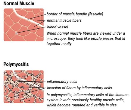
Drawing of Biopsy Showing Muscle Fibers Invaded by Immune Cells. Courtesy MDA.org

What Is Polymyositis?

[dropcap]P[/dropcap]olymyositis is a body-wide connective tissue disease resulting from autoimmune attack of skeletal muscles that is characterized by inflammatory and degeneratory changes. The course is unpredictable being marked by spontaneous flare-ups and remissions.

Polymyositis can begin slowly or abruptly according to the factor that is triggering the onset such as infection, medications like phenytoin, and autoimmune disease.

Progressive muscle weakness starts in the proximal skeletal muscles (muscles closest to the trunk of the body).

Skeletal muscles, also called voluntary, are muscles that move the body as we want, such as walking and  lifting objects, as opposed to those we cannot voluntarily control, such as the muscles of digestion. 

Q: What are the degeneratory changes in skeletal muscles?

A: In polymyositis, degeneratory changes in skeletal muscles means that muscles are being destroyed (called necrosis), resulting in fibrosis, or scarring. When scar tissue takes the place of lost muscle tissue, it cannot act like muscle to contract and relax.   Muscle destruction is what causes muscle pain and weakness.

After the clinical work-up of exams and blood studies to determine muscle damage, the diagnosis of polymyositis is confirmed by muscle biopsy. See image at above left. The black dots are inflammatory cells. Edema (fluid) between cells caused by inflammation pushes muscle fibers apart.

There is no cure for polymyositis, but the symptoms can be treated.  Options include medication, physical therapy, exercise, heat therapy (including microwave and ultrasound), orthotics and assistive devices, and rest.  The standard treatment for polymyositis is a corticosteroid drug, given either in pill form or intravenously.  Immunosuppressant drugs, such as azathioprine and methotrexate, may reduce inflammation in people who do not respond well to prednisone. 

Periodic treatment using intravenous immunoglobulin can also improve recovery.  Other immunosuppressive agents used to treat the inflammation associated with polymyositis include cyclosporine A, cyclophosphamide, and tacrolimus.  Physical therapy is usually recommended to prevent muscle atrophy and to regain muscle strength and range of motion.1

Diagnosis is based on elevated muscles enzymes, increased urinary creatine level, and electromyograph abnormalities.

Polymyositis can affect people at any age. It is most common in adults between ages 50 and 70, and in children ages 5 to 15. It affects women twice as often as men and is more common in African Americans than Caucasians.2 The major causes of death from polymyositis are cancer and lung disease, including pneumonia.  The 5-year mortality rate can be as high as 1 in 5 patients.2

What Is Polymyositis In Celiac Disease and/or Gluten Sensitivity?

Sources:
  1. National Institute of Neurological Disorders and Stroke []
  2. http://www.nlm.nih.gov/medlineplus/ency/article/000428.htm [] []

Scleroderma (Systemic Sclerosis)

sclerodermaWhat Is Scleroderma?

[dropcap]S[/dropcap]cleroderma is a chronic skin manifestation of progressive systemic sclerosis characterized by generalized thickened, edematous skin firmly bound to subcutaneous tissue which causes limited movement.

Systemic sclerosis a connective tissue disease that involves destructive changes in the skin, blood vessels, muscles, and internal organs. The course can be mild or it can be fatal. Cardiopulmonary complications from fibrosis are the most common cause of death.

Gastrointestinal problems mainly due to fibrosis affect 50 to 90% of patients.1

Q: Is there a cure for scleroderma?

A: There is no cure for scleroderma. Treatment is aimed at improving symptoms.

  • Heartburn (acid reflux) can be treated with antacid drugs.
  • Scleroderma kidney disease can be treated with blood pressure medications called “angiotensin converting enzyme inhibitors” (ACE inhibitors). These can often effectively control kidney damage if started early and use of these drugs has been a major advance for treating scleroderma.
  • Muscle pain and weakness can be treated with anti-inflammatory drugs such as prednisone, intravenous immunoglobin (IVIg), and/or immunosuppressive medications. Physical therapy may be useful to maintain joint and skin flexibility.2


What Is Scleroderma In Celiac Disease and/or Gluten Sensitivity?

Sources:
  1. Forbes A, Marie I. Gastrointestinal complications: the most frequent internal complications of systemic sclerosis. Rheumatology (Oxford). 2009 Jun;48 Suppl 3:iii36-9. doi: 10.1093/rheumatology/ ken485. []
  2. http://www.rheumatology.org []

Glycogenic Acanthosis (Esophageal Plaques)

15553
View of Esophagus Showing Distinctive Plaques In Glycogenic Acanthosis.

What Is Glycogenic Acanthosis?

Glycogenic acanthosis is a benign thickening of the esophageal squamous epithelium (surface cell lining)  characterized by the presence of numerous, uniformly grey-white plaques made up of large squamous cells filled with glycogen.

The wax-like plaques in glycogenic acanthosis are usually 2-10 mm in diameter and may be confluent round elevations involving the entire esophageal surface.1

 in radiographs (side-by-side) showing appearances of esphagus with glycogenic acanthosis. Courtesy The Radiology Assistant.com
Two radiographs (side-by-side) showing appearances of different esophagus with glycogenic acanthosis. Courtesy Radiology Assistant.nl

On x-ray  views of the well-distended esophagus, the plaques appear as a finely nodular or cobblestone mucosal pattern.

The findings are not associated with mucosal ulcerations, luminal narrowing, or mobility disturbance, although some patients may have coexistent hiatal hernia and gastroesophageal reflux.2

What Is Glycogenic Acanthosis In Celiac Disease and/or Gluten Sensitivity?

Sources:
  1. Suoglu OD, Emiroglu HH, Sokucu S, Cantez S, Cevikbas U, Saner G. Celiac disease and glycogenic acanthosis: a new association? Acta Paediatrica. Apr 2004;93(4):568-70. []
  2. Ghahremani GG, Rushovich AM. Glycogenic acanthosis of the esophagus: radiographic and pathologic features. Gastrointest Radiol. 1984;9(2):93-8. []

Dysphagia (Difficulty Swallowing)

What Is Dysphagia? [dropcap]D[/dropcap]ysphagia is a functional upper digestive disorder characterized by difficulty in swallowing or food getting stuck and swallowing that takes longer than 10 seconds to complete. Q: What determines the symptoms? A:… 

Post-Cricoid Carcinoma: definition

A malignancy with early metastasis and poor prognosis arising in the hypopharynx, the region of the throat lying behind the voice box (larynx). Symptoms start out as difficulty swallowing (dysphagia) that progress to the feeling…