This is a quote that a lot of people are hearing these days. What happens after your doctor says these words? Here is a synopsis of my journey; if any of this sounds familiar, give it some serious thought…there may be a very simple answer.
I first heard these words while I was barely concious, in a bed at Methodist Hospital in Houston. I had become so weak and debilitated by my undiagnosed and untreated celiac disease that my life was in danger. The symptoms over twenty years included gastrointestinal problems, fibromyalgia, irritability, bone pain and more. The good news was that I finally found out what was wrong with me; the better news was finding out that the disease is completely manageable through dietary changes alone.–no medications, no surgeries, none of that stuff. Just don’t eat grains that contain the gluten protein, mainly wheat, barley and rye. That sounded really good to me.
Not so fast. While still in the hospital, I was really still very sick, and not really able to process the information. After a couple of days (in which I was already improving), it dawned on me that
Calcium is the most abundant mineral in the body, with 99% residing in teeth and bones where it constitutes 40% of skeletal bone weight along with 45% phosphorus.
As a component of hard tissues, calcium fulfills a structural role to maintain body size and acts as attachments for musculoskeletal tissues.
Q: What does the non-skeleton calcium do in the body?
A: The remaining 1% of calcium is present in blood and soft tissues. Calcium levels in the blood are maintained within very strict limits by dietary intake, hormonal regulation and a rapidly exchangeable pool in bone tissue. The many important functions are described below.
What Is Calcium Deficiency In Celiac Disease and/or Gluten Sensitivity?
Woman with long standing osteoporosis. Courtesy of Wikimedia.
What Is Osteoporosis?
Osteoporosis is a metabolic bone disorder characterized by diminished bone mass (density) with normal cell appearance but fragile bone strength that prediposes to broken bones, and with high bone turnover.
This condition usually goes undetected until late when loss of height or a bone fracture occurs. In fact, each year 1.5 million fractures mainly of the hip, spine and wrist are attributed to osteoporosis. Compression fractures of vertebrae bones are the most common, accounting for 700,000 cases.
Bone is composed of specialized connective tissue called osseous tissue. Osseous tissue is made up of living bone cells (osteocytes) that are embedded in a hard matrix (framework) of calcified substance.
Bone matrix contains collagen fibers and the minerals calcium phosphate and calcium carbonate, which provide strength to bone. The copper enzyme, lysyl oxidase, is involved in the cross-linking of collagen in forming the framework for depositing calcium and other minerals to build and repair bone.
Q: How do osteocytes function in bone?
A: Osteocytes maintain the health of bone by their metabolic activity in regulating normal bone turnover. Bone turnover is the breaking down and removal of old or damaged bone and rebuilding or remodeling of healthy bone that is ongoing throughout life. The bone formation process takes about 3 months to complete.
Osteoporosis develops from failure of the body to maintain health and to provide bone tissue with adequate nutrition for proper function. Risk factors that can be modified include: low calcium intake, sedentary lifestyle, smoking, drinking alcohol excessively, eating a diet with excessive caffeine, protein, and phosphate, and taking certain medications over a long time such as steroids, thyroid preparations, the anti-convulsive drug phenytoin, aspirin, antacids, anticoagulants, some diuretics, and some chemotherapeutic drugs. See below for a fuller description.
In addition to celiac disease, osteoporosis is associated with advancing age, family history, nulliparity (no pregnancies) and post-menopause in females, certain disorders such as hyperthyroidism, hypogonadism, inflammatory bowel disease like Crohn’s disease, multiple myeloma, anorexia nervosa, and Cushing’s disease.
Bone strength is easily measured by testing bone mineral density (BMD). BMD is evaluated by DEXA scan (dual-energy X-ray absorptiometry). DEXA at the femoral neck and lumbar spine is considered the gold standard to confirm the diagnosis of osteoporosis. Results are expressed as T and Z scores. T scores compare the result with a 20 to 40 year old helathy person while Z scores compare the result with persons in the same age group. Both are measured in standard deviations (SD).
According to WHO criteria (World Health Organization), a T-score of -1 SD or greater denotes normal bone, a T-score between −1 to −2.5 SD denotes osteopenia, and a T-score of −2.5 or more denotes osteoporosis.1
Treatment is aimed to preserve and increase bone density, minimize symptoms for better quality of life and reduce risk of bone fractures.
What Is Osteoporosis In Celiac Disease and/or Gluten Sensitivity?
The cumulative effects of gluten-induced inflammation, treatment delay, and malabsorption result in lower bone density and bone fragility.2
Sources:
Pantaleoni S, Luchino M, Adriani A, Pellicano R, Stradella D, Ribaldone DG, Sapone N, Isaia GC, Di Stefano M, Astegiano M. Bone mineral density at diagnosis of celiac disease and after 1 year of gluten-free diet. ScientificWorldJournal. 2014;2014:173082. doi: 10.1155/2014/173082. [↩]
Grace-Farfaglia P. Bones of Contention: Bone Mineral Density Recovery in Celiac Disease-A Systematic Review. Nutrients. 2015 May 7;7(5):3347-3369. [↩]
Vitamin D is the principle regulator of calcium homeostasis (balance) in the body. This “vitamin” is really a prohormone, meaning it acts like a hormone but is not. Vitamin D does, however, contain cholesterol in its molecular structure like steroid hormones.
The physiological importance of vitamin D encompasses much more than the regulation of bone metabolism although this is a mighty function.
Q: How does vitamin D regulate bone metabolism?
A: In regulation of bone metabolism, vitamin D works in three ways: 1) enables active absorption of calcium from the small intestine, 2) enhances reabsortion of calcium by the kidneys that would otherwise be excreted in urine, and 3) plays an active role in skeletal development and bone mineralization. Mineralization gives strength to living bone tissue.
Vitamin D interacts with receptors within cells to effect transcriptional changes in many cell types including those in gut, bone, breast, prostate, brain, skeletal muscle, and the immune system.1
In regards to the essential role of vitamin D in muscle tissue, it has been recently shown that vitamin D regulates both muscle function and structure of primary myofibers.2
Vitamin D is converted in the body to a molecule that is biologically active. The active form is 1,25-dihydroxyvitamin D, usually referred to as vitamin D3. About 80% comes from sun exposure and the remaining from food.
Vitamin D3 is synthesized in the skin from 7-dehydrocholesterol via photochemical reactions requiring UV light (sunlight). That is, light that contains energy from the sun is incorporated into molecules of 7-dehydrocholesterol in the underlying dermis of skin to make this vitamin. This is why inadequate exposure to sunlight contributes to vitamin D deficiency.
Blood concentration of 25(OH)D is the best indicator of vitamin D status. It reflects vitamin D produced in the skin and that obtained from food and supplements and has a fairly long circulating half-life of 15 days.3
What Is Vitamin D Deficiency In Celiac Disease and/or Gluten Sensitivity?
Sources:
McCarty DE, Chesson AL Jr, Jain SK, Marino AA. The link between vitamin D metabolism and sleep medicine. Sleep Med Rev. 2014 Aug;18(4):311-9. doi: 10.1016/j.smrv.2013.07.001. [↩]
Girgis CM, Mokbel N, Cha KM, Houweling PJ, Abboud M, Fraser DR, Mason RS, Clifton-Bligh RJ, Gunton JE. The vitamin D receptor (VDR) is expressed in skeletal muscle of male mice and modulates 25-hydroxyvitamin D (25OHD) uptake in myofibers. Endocrinology. 2014 Sep;155(9):3227-37 [↩]
Potassium is a mineral that is crucial for life being essential for every cell, especially nerve and muscle function.
Most potassium is intracellular, meaning it is found within cells while sodium, its opposing mineral (both electrolytes), is found in the fluid surrounding cells.
In muscle contraction, exchange of potassium and sodium takes place so that potassium moves out of muscle cells and sodium moves into them.
With muscle relaxation, potassium moves back into the cells and sodium moves out. Functions are described below.
Importantly, a recent study investigating the association between the metabolic syndrome and potassium intake in the general population found a significant inverse association between potassium intake and metabolic syndrome in adults. That is, the lower the potassium intake, the greater the odds of developing metabolic syndrome. After adjusting for various lifestyle and dietary confounders, subjects in the highest quartile of potassium intake had 39% lower odds for metabolic syndrome compared to those in the lowest quartile. This association was consistent for both sexes. Among the components of metabolic syndrome, potassium intake was inversely related to abdominal obesity and fasting hyperglycemia in multivariate analysis.1
What Is Potassium Deficiency In Celiac Disease and/or Gluten Sensitivity?
Sources:
Shin D, Joh HK, Kim KH, Park SM. Benefits of potassium intake on metabolic syndrome: The fourth Korean National Health and Nutrition Examination Survey (KNHANES IV). Atherosclerosis. 2013 Sep;230(1):80-5. doi: 10.1016/j.atherosclerosis.2013.06.025. [↩]
Proptosis and Lid Retraction are Features of Grave’s Disease, or Hyperthyroidism.
What Is Grave’s Disease (Hyperthyroidism)?
G rave’s disease, or hyperthyroidism, is an autoimmune thyroid disease characterized by diffuse nontender goiter, elevated thyroxine hormone levels (T4, T3), suppressed thyroid stimulating hormone (TSH), and presence of thyroid receptor antibodies in the blood.
The autoantibodies involved are anti-thyroid peroxidase and anti-thyroglobulin antibodies. They bind to the thyroid stimulating hormone receptors, causing thyroid stimulation. These antibodies are detected by blood tests.
Q: What happens to the thyroid gland in Grave’s disease?
A: The thyroid gland is located in the front of the neck. This butterfly shaped gland consists of a large number of closed vesicles that contain a homogenous substance called colloid, which contains the thyroglobulin. Thyroglobulin is an iodine-containing protein secreted by the thyroid gland and stored within its colloid, from which the thyroid hormones thyroxine (T4) and triiodothyroinine (T3) are derived.1
Thyroxine molecule. Atoms are represented as spheres with conventional color coding: hydrogen (white), carbon (grey), oxygen (red), nitrogen (blue), iodine (purple).
T3 is the active hormone and is made from T4. Thyroid hormones are released into the bloodstream as needed to control metabolism, brain development, breathing, heart and nervous system functions, body temperature, muscle strength, skin dryness, menstrual cycles, weight, and cholesterol levels.
Thyroid hormone production is regulated by thyroid-stimulating hormone (TSH), which is made by the pituitary gland in the brain. Normally, when thyroid hormone levels in the blood are low, the pituitary releases more TSH in response to stimulation by the nearby hypothalamus which is continually monitoring levels of thyroxin. When thyroid hormone levels are high, the pituitary decreases TSH production. So in Grave’s disease, release of TSH by the pituitary gland is suppressed by the hypothalamus because thyroid hormone is elevated. Goiter develops from growth stimulation by thyroid stimulating autoantibodies.
What Is Grave’s Disease In Celiac Disease and/or Gluten Sensitivity?
Sources:
Taber’s Cyclopedic Medical Dictionary. 19th ed. F.A. Davis Company. Philadelphia, PA. [↩]
S eborrhea dermatitis is a recurring inflammatory disorder of sebaceous glands characterized by scaly patches of skin, often with bumps.
Seborrhea dermatitis results from the body’s inflammatory reaction to invasion by pityrosporum yeast that naturally inhabits the scalp and skin.1 Inflammation is the normal response to tissue injury and germ invasion.
Pityrosporum is a yeast that is commonly present worldwide. Its development depends on various factors that predispose to pityriasis versicolor, a chronic and mild superficial yeast infection. These infections usually are asymptomatic without itching or pain and without cellular and/or antibody responses.2
Q: Why are the sebaceous glands particularly affected by this yeast?
A: Pityrosporum yeast is an organism that needs oil produced by sebaceous glands to grow. If conditions permit, this yeast invades the superficial layer of skin and hair shafts to reproduce, causing infection. Such conditions include weakened skin due to nutritional deficiencies, excessive build-up of oil on skin, and altered immunity due to systemic disease such as psoriasis.
In adults, areas of skin that are the most affected have the greatest number of sebaceous glands especially the scalp, back, underarms, and the face including the eyelids, eyebrows and side folds of the nose.
All ages are subject to seborrhea dermatitis, and males have a higher occurence than females.
What Is Seborrhea In Celiac Disease and/or Gluten Sensitivity?
Zarei-Mahmoudabadi A, Zarrin. M, Mehdinezhad F. Seborrheic dermatitis due to Malassezia species in Ahvaz, Iran. Iran J Microbiol. 2013 Sep;5(3):268-71. [↩]
H ypertransaminasemia is a chronic condition of elevated blood liver transaminase enzymes, commonly called “liver enzymes,” that signifies hepatocellular (liver) injury.
Q: What are serum transaminases?
A: Transaminases are the liver enzymes ALT and AST. ALT is the abbreviation for alanine aminotransferase enzyme and AST is the abbreviation for aspartate aminotransferase enzyme.
Transaminases are commonly measured in routine blood tests to determine liver function. Elevated ALT and AST transaminases indicate inflammation of the liver. Other specific tests must follow to determine the cause of inflammation.
What Is Hypertransaminasemia In Celiac Disease and/or Gluten Sensitivity?
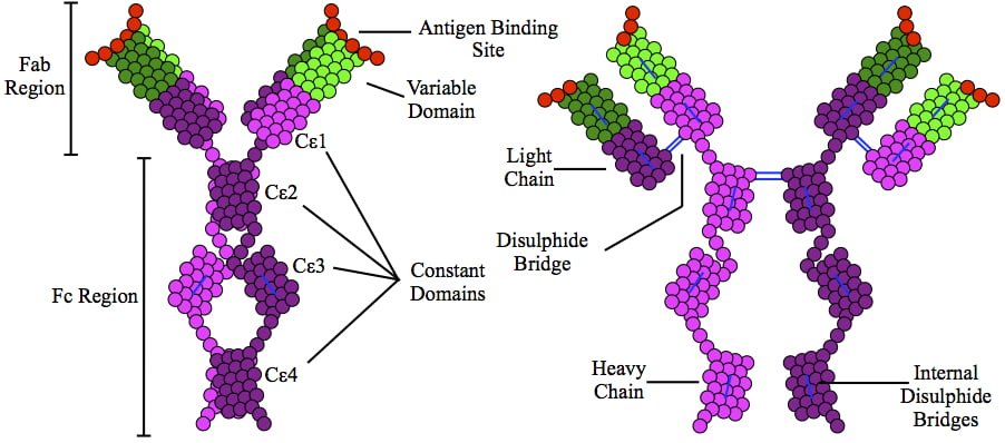
This is a depiction of immunoglobulin E (IgE) antibody which is elevated in allergic reactions.
What Is Allergic Rhinitis?
A llergic rhinitis is an immune disorder characterized by inflammation of the nasal mucosa by an IgE antibody reaction to an allergen.
An allergen is something that triggers an allergic immune response.
Q: What is the immune response?
A: Implicated in the response is an increase in T gamma-delta intraepithelial lymphocytes (IELs), which is a subset of pro-inflammatory T-cells located in the respiratory mucosa. Lymphocytes are white blood cells.
When a person with allergic rhinitis breathes in an allergen such as pollen or dust, the body releases chemicals, including histamine that cause allergy symptoms. For example, hay fever involves an allergic reaction to pollen. A similar reaction occurs with allergy to mold, animal dander, dust, and other allergens that are breathed in.
What Is Allergic Rhinitis In Celiac Disease and/or Gluten Sensitivity?
Deep Vein Thrombosis in the Right Leg with Swelling and Redness. Courtesy wikipedia.
What Is Antiphospholipid Syndrome?
Antiphospholipid syndrome (APS) is an autoimmune disease and a blood clotting disorder characterized by these clinical and laboratory criteria:
Clinical criteria – recurrent vascular thrombosis (clots in veins/arteries) from hypercoagulability (abnormal excessive clotting) and/or recurrent complications of pregnancy that include loss of the fetus (miscarriage) and pre-eclampsia or eclampsia.
Laboratory criteria – persistently elevated anticardiolipin, anti–beta-2 glycoprotein I, and/or lupus anti-coagulant antibodies in blood.
In antiphospholipid syndrome autoantibodies are produced by the body and directed against negatively charged phospholipids that are found in the outer layer of cell membranes and platelets. B2-glycoprotein-I (a protein in blood plasma) has been found as a major target antigen for antiphospholipid antibodies.
Q: Are phospholipids important in the body?
A: Yes. Phospholipid molecules are an essential part of cell membranes. They form a barrier around cells that protect the cell, allow movement of oxygen in and carbon dioxide out of the cell, and regulate other small molecules through the cell wall. Because phospholipids are widespread in the body, this disorder can produce a large variety of symptoms and affect many organs.
One severe effect of APS is the development of a blood clot in a vein deep in the arm or leg, called deep vein thrombosis (DVT). DVT can cause pain, swelling, redness, or increased warmth in the affected limb. Deep vein clots can break off, travel to the lungs, and cause pulmonary embolism.1 Pulmonary embolism is a medical emergency.
Treatment is with anticoagulant medications and blood monitoring.
What Is Antiphospholipid Syndrome In Celiac Disease and/or Gluten Sensitivity?