Skip to content

DHA (Docosahexaenoic Acid) Deficiency

Smoked Salmon...Brain Food that's Good for the Eyes!
Salmon…Brain Food That’s Good for Your Eyes, Heart, Blood Vessels, Blood Sugar, Liver, Muscles, and Fights Inflammation!

What Is DHA?

[dropcap]D[/dropcap]ocosahexaenoic acid (DHA) is an essential omega-3 fatty acid that is abundant in the brain, being crucial in brain structure. As such DHA is a key component of neuronal membranes together with arachidonic acid (a major opposing omega-6 fatty acid), making up 15-20% of the brain’s dry mass.

This polyunsaturated fatty acid is obtained from fish sources of food.

In healthy human volunteers, positron emission tomography (PET) has shown that the normal human brain consumes 4.6 mg/day of DHA.1

DHA is particularly concentrated in highly active membranes such as nerve synapses (junctions) and photoreceptors in the eye (retina).

Q: How much DHA is in the retina?

A: DHA makes up more than 30% of the retina.2

In other roles, DHA is an  important building material for the eicosanoids, a large group of highly bioactive hormone-like substances including prostaglandins, leukotrienes, and thromboxanes that are involved in blood clotting, inflammation, and vasoconstriction.

DHA has been shown to increase insulin sensitivity as opposed to the opposite problem of insulin resistance, to improve muscle mass, and protect against non-alcoholic fatty liver disease.3

Egert et al. in a study of people aged 19 to 43 years with normal cholesterol showed that DHA intake significantly increased serum HDL (good) cholesterol. Also, DHA significantly decreased fasting serum triglycerides.4

What Is DHA Deficiency In Celiac Disease and/or Gluten Sensitivity?

Sources:
  1. Rapoport SI. Brain arachidonic and docosahexaenoic acid cascades are selectively altered by drugs, diet and disease. Prostaglandins Leukot Essent Fatty Acids. 2008 Sep-Nov;79(3-5):153-6. Epub 2008 Oct 29. []
  2. Richardson AJ. The importance of omega-3 fatty acids for behavior, cognition, and mood. Scandinavian Journal of Nutrition. 2003;47(2):92-8 []
  3. Espinosa A, Valenzuela R, González-Mañán D, D’Espessailles A, Guillermo Gormaz J, Barrera C, Tapia G. Prevention of liver steatosis through fish oil supplementation: correlation of oxidative stress with insulin resistance and liver fatty acid content. Arch Latinoam Nutr. 2013 Mar;63(1):29-36. []
  4. Egert S, Kannenberg F, Somoza V, et al. Dietary alpha-linolenic acid, EPA, and DHA have differential effects on LDL fatty acid composition but similar effects on serum lipid profiles in normolipidemic humans. J Nutr. 2009;139:861–868. doi: 10.3945/jn.108.103861 []

Multiple Sclerosis

Brain scan showing changes in multiple sclerosis over a month. Courtesy Wikimedia
Brain scan showing changes in multiple sclerosis by month over a year’s time. Courtesy Wikimedia

What Is Multiple Sclerosis?

[dropcap]M[/dropcap]ultiple sclerosis is an autoimmune disease of the central nervous system characterized by patchy inflammation of the myelin sheath surrounding nerve cells that produces multiple and varied neurologic symptoms and signs due to demyelination.

Q: What does demyelination mean.

A: Demyelination means there is damage to the myelin sheath which is a fatty substance that surrounds and protects nerve cells much like a the covering of a lamp cord keeps the electricity flowing within it from the plug to the light bulb. This damage impairs transmission of nerve impulses between the brain and body.

In the process of demyelination, T-lymphocyte cells that are activated and potentially autoimmune cross the blood-brain barrier and produce inflammatory plaques and axonal tissue loss in the brain, spinal cord or optic (vision) nerves. The end result is the accumulation of gliosis (scarring) and demyelination of areas in the central nervous system.1

The course of multiple sclerosis is unpredictable and slowly progressive, usually with  exacerbations (worse symptoms) and remissions (relief of symptoms).

Multiple sclerosis affects about 1 ‰ of the population worldwide.

What Is Multiple Sclerosis In Celiac Disease and/or Gluten Sensitivity?

Sources:
  1. Rodrigo L, Hernández-Lahoz C, Fuentes D, Alvarez N, López-Vázquez A, González S. Prevalence of celiac disease in multiple sclerosis. BMC Neurol. 2011 Mar 7;11:31. doi: 10.1186/1471-2377-11-31. []

Vitamin A Deficiency

Mango Salad.
Mango Salad.

What Is Vitamin A?

[dropcap]V[/dropcap]itamin A is a family of fat-soluble compounds that are essential for health, vision, protection of the body, reproduction, and normal growth of children before and after birth.

Q: What is the family of vitamin A?

A: The natural vitamin A family members are called retinoids. Retinoids are found only in animal food sources. They include retinol (alcohol form), retinal (aldehyde form), and retinoic acid (acid form).

In the plant kingdom, pigments called carotenoids can yield retinoids on metabolism in the body. Carotenoids that can converted to retinol include β-carotene, α-carotene, and β-cryptoxanthin.1

There are hundreds of various carotenoids although only a few have been researched including beta-carotene, lycopene, lutein and zeaxanthin.

Fruits and vegetables that are a rich source of carotenoids are thought to provide health benefits by decreasing the risk of various diseases, particularly eye diseases and certain cancers (esophageal, stomach, pancreatic, bladder and cervical). In this respect, research shows that the regular consumption of fruits and vegetables may prevent 20% of most types of cancers.2

In part, the beneficial effects of carotenoids are thought to be due to their role as antioxidants. Lutein and zeaxanthin may be protective in eye disease because they absorb damaging blue light that enters the eye.3

The antioxidant effects appear to be highest in fresh fruit as compared to preserved fruit. A study of fresh peach pulps and peel in animals demonstrated highest antioxidant and anti-inflammatory effects in fresh fruits preventing against induced damage in animal tissues.4

A study investigating the specific anti-inflammatory and anti-oxidant micronutrients that reduce oxidative stress showed that adolescents with metabolic syndrome (MetS) had consistently lower carotenoid concentrations compared with their counterparts without MetS. MetS prevalence was estimated at 7% among boys and 3% among girls.5

What Is Vitamin A Deficiency In Celiac Disease and/or Gluten Sensitivity?

Sources:
  1. Maggio M, de Vita F, Lauretani F, et al. Relationship between Carotenoids, Retinol, and Estradiol Levels in Older Women. Nutrients. 2015 Aug 5;7(8):6506-19. doi: 10.3390/nu7085296. []
  2. Gasparotto J, Somensi N, Bortolin RC, Moresco KS, Girardi CS, Klafke K, Rabelo TK, Morrone Mda S, Vizzotto M, Raseira Mdo C, Moreira JC, Gelain DP. Effects of different products of peach (Prunus persica L. Batsch) from a variety developed in southern Brazil on oxidative stress and inflammatory parameters in vitro and ex vivo. J Clin Biochem Nutr. 2014 Sep;55(2):110-9. doi: 10.3164/jcbn.13-97. Epub 2014 Sep 1. []
  3. Krinsky NI1, Johnson EJ. Carotenoid actions and their relation to health and disease. ((Mol Aspects Med. 2005 Dec;26(6):459-516. []
  4. Gasparotto J, Somensi N, Bortolin RC, Moresco KS, Girardi CS, Klafke K, Rabelo TK, Morrone Mda S, Vizzotto M, Raseira Mdo C, Moreira JC, Gelain DP. Effects of different products of peach (Prunus persica L. Batsch) from a variety developed in southern Brazil on oxidative stress and inflammatory parameters in vitro and ex vivo. J Clin Biochem Nutr. 2014 Sep;55(2):110-9. doi: 10.3164/jcbn.13-97. []
  5. TGBeydoun MA, Canas JA, Beydoun HA, Chen X, Shroff MR, Zonderman AB. Serum antioxidant concentrations and metabolic syndrome are associated among U.S. adolescents in recent national surveys. J Nutr. 2012 Sep;142(9):1693-704. doi: 10.3945/jn.112.160416. []

Uveitis, Non-Infectious  

Drawing of eyeball to illustrate uveitis. Courtesy National Eye Institute
Drawing of Eye to Illustrate Uvea Tract. Courtesy
National Eye Institute

What Is Non-Infectious Uveitis?

[dropcap]N[/dropcap]on-infectious uveitis, as opposed to that which is caused by an infection, is an inflammatory eye disorder characterized mainly by swelling of the uveal tract structures in the anterior (front) of the eye.  

Inflammation can also affect the lens, retina, optic nerve, and vitreous, producing reduced vision or blindness if left untreated. 

Q: What are structures of the uveal tract?

A: Structures of the uveal tract are composed of the iris, the ciliary body, and the choroid: 

  • The iris is the colored circle at the front of the eye. It defines eye color, secretes nutrients to keep the lens healthy, and controls the amount of light that enters the eye by adjusting the size of the pupil (opening).
  • The ciliary body is located between the iris and the choroid. It helps the eye focus by controlling the shape of the lens and it provides nutrients to keep the lens healthy.
  • The choroid is a thin, spongy network of blood vessels, which primarily provides nutrients to the retina in the back of the eye.1

Uveitis may be caused by problems or diseases occurring in the eye itself or it can be part of an inflammatory disease affecting other parts of the body. The uveal tract has a rich supply of blood vessels that contain immune cells to fight microbial invasion. These immune cells are lymphocytes, phagocytes, and plasma cells.

The uvea can be attacked by autoimmune antibodies produced in autoimmune diseases that affect similar tissues in other parts of the body such as psoriasis, Behcet’s syndrome, multiple sclerosis, sarcoidosis, Vogt Koyanagi Harada’s disease, and celiac disease.

Uveitis can last for a short (acute) or a long (chronic) time. The severest forms of uveitis reoccur many times.

What Is Non-Infectious Uveitis In Celiac Disease and/or Gluten Sensitivity?

Sources:
  1. http://www.nei.nih.gov/health/uveitis/uveitis.asp []

Xerophthalmia

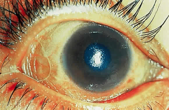
Marked conjunctival and corneal xerosis. The entire bulbar conjunctiva is dry and thickened and almost skinlike. (Sommer A, Sugana T, Djunaedi E, Green R: Vitamin A-responsive panocular xerophthalmia in a healthy adult. Arch Ophthalmol 96:1630, 1978)

What Is Xerophthalmia?

[dropcap]X[/dropcap]erophthalmia is a serious condition of extreme drying and thickening of the conjunctiva following chronic inflammation and failure to make tears, characterized by hazy, dry cornea.

Vitamin A is obtained from the diet and is required in the cornea for maintaining epithelial (surface cell) health.1

Xerophthalmia is most often associated with malnutrition involving chronic, severe vitamin A deficiency and may develop from chronic liver disease, intestinal disorders such as celiac disease, pancreatitis, and extensive bowel surgery that cause malabsorption.

It may also develop in autoimmune diseases such as Sjogrens syndrome.

Q: What is the prognosis for xerophthalmia?

A: Xerophthalmia has the potential to rapidly advance to corneal necrosis (keratomalacia) and blindness.

What Is Xerophthalmia In Celiac Disease and/or Gluten Sensitivity?

Sources:
  1. Alwitry A. Vitamin A deficiency in coeliac disease. Br J Ophthalmol. 2000 September; 84(9): 1075. doi:  10.1136/bjo.84.9.1075e []

Keratomalacia

 Keratomalacia in a severely malnourished 10-month-old Indonesian boy.  (Sommer A, Sugana T: Corneal xerophthalmia and keratomalacia. Arch Ophthalmol 100:404, 1982)
Keratomalacia in a severely malnourished 10-month-old Indonesian boy.1

What Is Keratomalacia?

[dropcap]K[/dropcap]eratomalacia is a severe non-inflammatory eye condition characterized by a hazy, dry cornea that becomes softened and denuded (loss of protective layer), which when liquified is then irreversibly destroying the eyeball and causing blindness.

The conjunctiva and lacrimal glands (tear producing) are also affected resulting in ulceration of the eye surface with, or without, Bitot’s spots (foamy patches) because these tissues also require adequate vitamin A to function normally.

The conjunctiva is the mucous membrane that lines the inside of the eyelids and covers the eyeball surface, also called the white of eye.

Q: What does the term keratomalcia mean?

A: In the term keratomalcia, kerato refers to the cornea and malacia means softening of the tissues.

What Is Keratomalacia In Celiac Disease and/or Gluten Sensitivity?

Sources:
  1. Sommer A, Sugana T: Corneal xerophthalmia and keratomalacia. Arch Ophthalmol 100:404, 1982 []

Occipital Lobe Epilepsy With Cerebral Calcifications

CT scan showing brain calcifications. Courtesy Wikimedia

What Is Occipital Lobe Epilepsy With Cerebral Calcifications?

[dropcap]O[/dropcap]ccipital lobe epilepsy with cerebral calcifications is a seizure disorder that develops from calcium deposits in the brain.

This condition is characterized by repetitive abnormal electrical discharges within the brain that may manifest as paroxysmal, or sudden recurrence of, visual disturbances.

Q: Where is the occipital lobe?

A: The occipital lobe is one of four lobes of the cerebrum, or upper brain. It comprises the lower rear portion of the cerebrum and mainly processes visual images coming to  it from the eyes through the optic nerve.

In this image of the left side of the brain, the occipital lobe is pink. From Wikimedia.                                                                                      Drawing shows the 4 lobes of the human brain on the left side. The occipital lobe is pink. Courtesy Wikimedia

What Is Occipital Lobe Epilepsy With Cerebral Calcifications In Celiac Disease and/or Gluten Sensitivity?