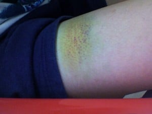
Editors’ note: This case report illustrates that a person can live a long time reporting apparent good health and be completely unaware that they have symptoms of celiac disease. In this case, hematomas, (which are swollen black and blue marks caused by a break in the wall of a blood vessel), that developed on his legs caused the patient to seek medical attention. The ability of his blood to clot was severely impaired and yet there was no other manifestation of hemorrhage. Discover more about bruising and hundreds of other health issues and how to treat them at the Gluten Free Works Treatment Guide.
Salmon…Brain Food That’s Good for Your Eyes, Heart, Blood Vessels, Blood Sugar, Liver, Muscles, and Fights Inflammation!
What Is DHA?
Docosahexaenoic acid (DHA) is an essential omega-3 fatty acid that is abundant in the brain, being crucial in brain structure. As such DHA is a key component of neuronal membranes together with arachidonic acid (a major opposing omega-6 fatty acid), making up 15-20% of the brain’s dry mass.
This polyunsaturated fatty acid is obtained from fish sources of food.
In healthy human volunteers, positron emission tomography (PET) has shown that the normal human brain consumes 4.6 mg/day of DHA.1
DHA is particularly concentrated in highly active membranes such as nerve synapses (junctions) and photoreceptors in the eye (retina).
In other roles, DHA is an important building material for the eicosanoids, a large group of highly bioactive hormone-like substances including prostaglandins, leukotrienes, and thromboxanes that are involved in blood clotting, inflammation, and vasoconstriction.
DHA has been shown to increase insulin sensitivity as opposed to the opposite problem of insulin resistance, to improve muscle mass, and protect against non-alcoholic fatty liver disease.3
Egert et al. in a study of people aged 19 to 43 years with normal cholesterol showed that DHA intake significantly increased serum HDL (good) cholesterol. Also, DHA significantly decreased fasting serum triglycerides.4
What Is DHA Deficiency In Celiac Disease and/or Gluten Sensitivity?
Sources:
Rapoport SI. Brain arachidonic and docosahexaenoic acid cascades are selectively altered by drugs, diet and disease. Prostaglandins Leukot Essent Fatty Acids. 2008 Sep-Nov;79(3-5):153-6. Epub 2008 Oct 29. [↩]
Richardson AJ. The importance of omega-3 fatty acids for behavior, cognition, and mood. Scandinavian Journal of Nutrition. 2003;47(2):92-8 [↩]
Espinosa A, Valenzuela R, González-Mañán D, D’Espessailles A, Guillermo Gormaz J, Barrera C, Tapia G. Prevention of liver steatosis through fish oil supplementation: correlation of oxidative stress with insulin resistance and liver fatty acid content. Arch Latinoam Nutr. 2013 Mar;63(1):29-36. [↩]
Egert S, Kannenberg F, Somoza V, et al. Dietary alpha-linolenic acid, EPA, and DHA have differential effects on LDL fatty acid composition but similar effects on serum lipid profiles in normolipidemic humans. J Nutr. 2009;139:861–868. doi: 10.3945/jn.108.103861 [↩]
Photo by gastroscopy showing ulcer in the antrum area of the stomach (lower area).
What Is A Gastric Ulcer?
Gastric ulcer is a painful stomach disorder characterized by an open sore involving the mucosa lining and deeper muscle layer of the stomach.
Gastric ulcer is associated with lymphocytic gastritis which is inflammation of the mucosal lining of the stomach. The thick mucosal lining normally protects the stomach from the erosive action of stomach acid.
Q: How do ulcers develop?
A: Ulcers develop if hydrochloric acid secreted by the gastric glands of the stomach for the purpose of digesting food damages the normally resistant mucosal walls of the stomach. In the reverse, ulcers may be accompanied by achlorhydria (insufficient acid production).
Damage occurs when there is a predisposing factor that alters the health of the mucosal lining. The most common cause is infection with a bacteria called h. pylori bacter, stress and chronic use of the pain relievers aspirin and non-steroidal drugs like ibuprofen.
Smoking tocacco and consuming alcohol aggravate an ulcer but do not cause it to develop.
The most common location for ulcer formation is along the stomach antrum which is the area of the stomach before the pylorus, the lower region that empties liquid stomach contents into the small intestine.
What Is A Gastric Ulcer In Celiac Disease and/or Gluten Sensitivity?
Testing the Eye for Tear Production (L) and Damage to Conjunctiva from Dryness (R).
What Is Sjögren’s Syndrome?
S jögren’s syndrome is a systemic inflammatory autoimmune disease with a chronic, progressive course that primarily attacks the lacrimal glands of the eye and the salivary glands of the mouth, which are exocrine glands. Exocrine glands secrete the substances they produce through a duct.
Sjögren’s syndrome is ordinarily characterized by dysfunction of the lacrimal glands to produce tears causing dry eye and the salivary glands to produce saliva causing dry mouth, but is not limited by or to these features.
Besides involvement of these exocrine glands, there may be involvement of other parts of the body, termed extraglandular, which may be more severe than eye or mouth features.
There is not yet agreement on classifying Sjögren’s syndrome. Primary and secondary are the two forms generally accepted.1 Both forms can cause mild to severe disease, called the spectrum:
Primary Sjögren syndrome. Disease occurs without involvement of other linked autoimmune disorders. In addition to the eyes and mouth, the nose, throat and skin may also be affected and joints, lungs, kidneys, blood vessels, digestive organs and nerves as well.2 Systemic manifestations (other than eyes and mouth) concern a third of patients, including lymphoma in 5% of the patients.3
Secondary Sjögren’s syndrome. Disease complicates other autoimmune disease such as systemic lupus erythematosus, rheumatoid arthritis, primary biliary cirrhosis, and celiac disease.
Diagnosis of Sjögren’s syndrome is made by most doctors based on Schimer’s test for tears and unstimulated whole salivary flow to assess objective eye and oral involvement, since these are the tests most physicians use in clinical practice.4 Specific antibody tests would be positive for anti-Ro (SSA)/anti-La (SSB) autoantibodies. Sjögren’s syndrome should also be considered when extraglandular manifestations such as vasculitis, polyneuropathy or arthritis occur, even when the patients do not complain of dry eyes and mouth.5
There is no cure for Sjögren’s syndrome. Treatment is aimed to diminish symptoms. For example, steroids and Ibupropen are used to decrease inflammation and pain in joints. Artificial tears and ointments are used for dry eye.
Most people who develop Sjogren’s syndrome are older than 40 years. Nine of ten people with Sjögren’s syndrome are women.2
What Is Sjögren’s Syndrome In Celiac Disease and/or Gluten Sensitivity?
Sources:
Huang YF, Cheng Q, Jiang CM, An S, Xiao L, Gou YC, Yu WJ, Lei L, Chen QM, Wang Y, Wang J. The immune factors involved in the pathogenesis, diagnosis, and treatment of Sjogren’s syndrome. Clin Dev Immunol. 2013;2013:160491. doi: 10.1155/2013/160491. Epub 2013 Jul 9. [↩]
Fazaa A, Bourcier T, Chatelus E, Sordet C, Theulin A, Sibilia J, Gottenberg JE. Classification criteria and treatment modalities in primary Sjögren’s syndrome. Expert Rev Clin Immunol. 2014 Apr;10(4):543-51. doi: 10.1586/1744666X.2014.897230. [↩]
Cornec D, Saraux A, Cochener B, Pers JO, Jousse-Joulin S, Renaudineau Y, Marhadour T, Devauchelle-Pensec V. Level of agreement between 2002 American-European Consensus Group and 2012 American College of Rheumatology classification criteria for Sjogren’s syndrome and reasons for discrepancies. Arthritis Res Ther. 2014 Mar 19;16(2):R74. [↩]
Witte T. Pathogenesis and diagnosis of Sjögren’s syndrome. Z Rheumatol. 2010 Feb;69(1):50-6. doi: 10.1007/s00393-009-0519-2. [↩]
What Are Erosions In The Second Part Of The Duodenum?
Erosions in the second part of the duodenum typically are superficial excoriations (sores) in the mucosal lining that do not penetrate into the muscle layer as does an ulcer.
Of note, most erosions develop in the first part of the duodenum rather than the second part.
Q: Are duodenal erosions serious?
A: Yes duodenal erosions are serious and can be life-threatening for the following reasons:
Duodenal erosions may occur together with gastric erosions, therefore they can jointly induce massive bleeding.
Duodenal erosions may be associated with duodenal ulcer but hemorrhage originates from the erosion and the ulcer itself does not bleed.
Duodenal erosion in itself might induce bleeding. The intensity of bleeding, if there is no hemorrhage from other places, is not so severe, as the one originating from gastric erosions.1
What Are Erosions In The Second Part Of The Duodenum In Celiac Disease and/or Gluten Sensitivity?
Sources:
Preisich P, Farkas I, Konyár EJ. The role of duodenal erosions in the occurrence of upper gastrointestinal haemorrhage. Scand J Gastroenterol Suppl. 1989;167:36-8. [↩]
Immune thrombocytopenia purpura (ITP), formerly called idiopathic, is an immune mediated bleeding disorder characterized by destruction of circulating platelets (thrombocytes) by autoantibodies in the presence of normal bone marrow and without other conditions that cause bleeding problems including many medications.
Q: How do autoantibodies destroy platelets?
A: Autoantibodies bind to normal platelet surface antigens (targets) which forms a complex that is cleared from blood by the reticuloendothelial system. Consequently, this condition results in an abnormal decrease in the number of platelets circulating in blood plasma.
Platelets are cells that are needed to stop bleeding. A normal platelet count is in the range of 150,000 to 450,000. With ITP, the platelet count is less than 100,000. By the time significant bleeding occurs, the patient may have a platelet count of less than 10,000. The lower the platelet count, the greater the risk of bleeding.
Because of inability of blood to properly clot due to lack of sufficient platelets, a distinctive purplish rash develops under the skin from blood leakage and an appearance of many small red spots of varying size can cover the skin. Depending on the number of platelets, there may be minimal to extensive bleeding in various parts of the body. Bleeding in the brain is the worst complication, causing death in approximately 1% of affected children and 5% of affected adults.
Immune thrombocytopenic purpura is common in childhood, on average 50 cases per 1,000,000 per year with same incidence in boys as girls. It may follow a viral infection or immunization and is caused by an inappropriate response of the immune system to cross react against the body’s platelets.1
The incidence of immune thrombocytopenic purpura in adults is approximately 66 cases per 1,000,000 per year. Older persons and persons with a previous history of hemorrhage are at increased risk of severe bleeding in adult ITP.
Spontaneous remission occurs in most children (80%). Remission in adults usually requires treatment.
What Is Immune Thrombocytopenic Purpura In Celiac Disease and/or Gluten Sensitivity?
Sources:
Dogan M, Sal E, Akbayram S, Peker E, Cesur Y, Oner AF. Concurrent celiac disease, idiopathic thrombocytopenic purpura and autoimmune thyroiditis. Clin Appl Thromb Hemost. 2011 Nov-Dec;17(6):E13-6. doi: 10.1177/1076029610378502. Epub 2010 Aug 3. [↩]
Transient erythroblastopenia is a rare disorder of red blood cell formation characterized by brief, reversible disappearance of erythroblasts (red blood cell precursors) in the bone marrow of children.
Q: What do the red blood cells look like?
A: Circulating red blood cells appear normal so that the anemia which develops is a normocytic (normal cellular) anemia in contrast to microcytic (small cell) anemia in iron deficiency or macrocytic (large cell) anemia in vitamin B12 or folic acid deficiency.
Because new red blood cells are failing to mature, packed red blood cell transfusions may be necessary in severe anemia.
In adults, this disorder is called pure red blood cell anemia.
What Is Transient Erythroblastopenia In Celiac Disease and/or Gluten Sensitivity?
Cancel your subscription?
We're sorry to see you go.
Please select a reason for cancellation.
Please provide additional details.
Cancellation request submitted
We've received your request. Your subscription remains active while we process it. We'll email you once it's confirmed.
Cancellation pending
Access Content
Affiliate Terms
By joining our Affiliate Program, you agree to the following terms:
1. Eligibility: You must be 18 years or older to participate. We reserve the right to refuse service to anyone for any reason at any time.
2. Promotional Guidelines: Affiliates must not engage in misleading advertising or spam. Promotions must accurately represent our brand and products.
3. Affiliate Links: You must use your unique tracking link to receive credit for referred sales.
4. Prohibited Use: You may not use our branding in a way that may confuse customers or misrepresent your relationship with us.
5. Termination: We reserve the right to terminate your account at any time for breach of these terms.
6. Changes to Agreement: We may update these terms at any time. Continued participation implies acceptance of any changes.
By continuing, you agree to the terms of this Affiliate Agreement.