[dropcap]A utoimmune disorders refer to those conditions that involve an abnormal attack on the body’s own tissues perpetuated by the production of autoantibodies directed against self.
Q: What happens when autoantibodies attack the body’s own tissues?
A: This abnormal immune activity by autoantibodies causes inflammation and damage to targeted body tissues.
Dermatitis herpetiformis is a skin manifestation of celiac disease characterized by extremely itchy blisters that commonly erupt on forearms and knees but may appear on the face, scalp or buttocks.
Autoimmune diseases as a group affect approximately 8.5% of people worldwide.
What Are Autoimmune Disorders In Dermatitis Herpetiformis?
Microscopic view of pancreatic islet cells. Courtesy Dr. José Sánchez Gonzales
What Is Type I Diabetes Mellitus?
[dropcap]T ype 1 diabetes mellitus (T1DM), also termed type 1A, is an inherited autoimmune disorder in which anti-islet autoantibodies destroy the islet cells of the pancreas that secrete insulin hormone. Type 1 diabetes mellitus was formerly called juvenile diabetes because it usually afflicts persons under the age of 25 years.
Loss of insulin production results in failure to metabolize glucose. Glucose is a simple sugar that is a required source of energy for the body, especially the brain and muscles.
Type 1 diabetes mellitus is characterized by sustained fasting blood glucose levels above 126 mg/dL (hyperglycemia) with subsequent loss of glucose from the body by removal through the urine (glucosuria) as the body attempts to lower blood glucose, and cell starvation that follows.
That is, while glucose accumulates in blood, the body cannot access it. Without insulin treatment, this disorder quickly produces coma and ultimately results in death. In fact, it is 5th leading cause of death in the United States.
Q: How does insulin work?
A: Insulin moves glucose from the bloodstream into body cells where it is used or reformulated for high energy storage. For example, muscles can use glucose for immediate work or store it in the form of glygogen for later work, depending on need. Healthy insulin production keeps an 8 hour fasting blood glucose level to less than 100 mg/dL. Upon eating carbohydrate food, glucose is digested and absorbed from the small intestine into the bloodstream which then raises blood glucose levels. The elevated level is controlled by prompt action of insulin to lower it to below 140 mg/dL within 2 hours of eating.
Insulin does not work alone. The islets of Langerhans manage glucose in the body. The islets are specialized formations located on the outer surface of the pancreas. The islets are composed of two different types of cells known as alpha and beta cells. These cells make the competing hormones that keep blood glucose within a healthy range.
Alpha cells secrete glucagon to raise blood glucose levels by triggering the body to release stored energy in the form of glycogen. In the opposite, beta cells secrete insulin to lower blood glucose by opening body cells so that glucose in blood can enter. Without insulin, glucose cannot enter cells but remains in the bloodstream where it accumulates.
Insulin is also needed to move magnesium into cells from the bloodstream. On the other side, magnesium is needed to produce insulin. Insulin has other functions such as building muscle and helping regulate cholesterol which directly impacts the sex hormones, estrogen, progesterone, and testosterone.
Onset of symptoms usually occurs over a period of days or weeks, although beta cell destruction can begin years earlier. The SEARCH for Diabetes in Youth multicenter study, funded by the Centers for Disease Control and Prevention (CDC) and the National Institutes of Health (NIH), has determined that based on data from 2002 to 2003, a total of 15,000 youth in the United States were newly diagnosed with type 1 diabetes each year. Non-Hispanic white youth had the highest rate of new cases of type 1 diabetes according to NIH.
Type 1A diabetes mellitus has become one of the most intensively studied autoimmune disorders. It is now possible to predict its development, beginning with HLA-encoded genetic susceptibility, followed by the development of a series of anti-islet autoantibodies.1
What Is Type I Diabetes Mellitus In Celiac Disease and/or Gluten Sensitivity?
Sources:
Liu E, Eisenbarth GS. Type 1A diabetes mellitus-associated autoimmunity. Endocrinology and Metabolism Clinics of North America. Jun 2002;31(2):391-410, vii-viii. [↩]
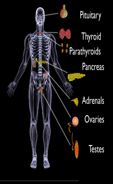
Endocrine glands targeted in polyglandular autoimmune syndrome.
What Are Autoimmune Polyglandular Syndromes?
[dropcap]A utoimmune polyglandular syndromes (APS) are rare clusterings of two or more endocrine and non-endocrine autoimmune disorders in the same affected person.
Polyglandular is somewhat of a misnomer since many of the manifestations of the diseases do not concern endocrine glands.1
Endocrine autoimmune disorders involve the abnormal production of autoantibodies that target and destroy the body’s own endocrine tissues, causing loss of essential hormone production by the targeted glands. Endocrine glands include the pituitary, thyroid, adrenal, parathyroid, islets of Langerhans (pancreas), testes in males, and ovaries in females.
First degree relatives (siblings of same parents, parents, children) have an increased incidence of latent, meaning not apparent, autoimmune pathology.2
Q: How many autoimmune polyglandular syndromes are described?
A:Three syndromes have been identified and they are all inherited: APS type-1, APS type-2, and APS type-3.
APS type-1 is a genetic mutation inherited in an autosomal recessive manner. A child with APS type-1 has inherited two mutated copies of a gene called the AIRE (autoimmune regulator) gene, which is on the long arm of 21st chromosome present in each cell.3 The parents, called carriers, are unaffected since they each have only a single copy of the AIRE mutated gene. Humans have a total of 23 pairs of chromosomes that contain genes inherited from each parent. Mutations in the genes cause disease.
Diagnosis criteria for autoimmune polyglandularsyndrome type-1 includes these three disorders:
Chronic candida infection (CMC), which usually develops first, typically attacks skin, but very commonly also nails, mouth, vagina, esophagus and intestine. CMC in APS type-1 patients is usually mild, and in most cases, it is chronic. It is found in 73–100 % of APS type-1 patients.
Hypoparathyroidism, causing loss of parathyroid function (hypoparathyreosis) is found in 76–93 % of APS type-1 patients.
Autommune Addison’s disease, also called autoimmune adrenalitis, is found in 72-100 % of APS type-1 patients. Still many of them die for unrecognized or late diagnosed autoimmune Addison’s disease, so regular follow-up for children in suspicion of APS type-1 (with CMC or/and hypoparathyroidism) is necessary.4
Other assocated disorders that may develop, but are not required for diagnosis, include: vitiligo, premature menopause, pernicious anemia, parathyroid gland failure, alopecia, and celiac disease. Thyroid disease rarely occurs.5
APS type-2 is linked to the inheritance of HLA antigens on chromosome 6 and appears to be autosomal dominant with incomplete penetrance. This suggests the contribution of environmental factors, such as bacterial and viral infections, medications, psychological factors, etc.6It does not have an identified mutation of the AIRE gene.
Diagnosis criteria for autoimmune polyglandular syndrome type-2 includes these two disorders:
Autommune Addison’s disease combined with
Autoimmune thyroid disease (thyroid atrophy, hypertrophic goiter related to Hashimoto’s thyroiditis, Graves’ disease, asymptomatic autoimmune thyroiditis)6 and/or type I diabetes mellitus. The conditions may occur in any order.
Polyglandular autoimmune syndrome type-2 is also known as Schmidt’s syndrome when adrenalitis (adrenal insufficiency) is associated with thyroiditis and Carpenter’s syndrome for adrenal insufficiency with hypoparathyreosis (impaired function of parathyroid glands).
Other disorders that may develop, but are not required for diagnosis, include: type 1 diabetes (50%), frequently gonadal failure or vitiligos, also celiac disease, autoimmune hepatitis, alopecia, pernicious anemia, and myasthenia gravis.7 Decades may arise between the onset of one disease and the onset of the second in the same patient.8
Therapy of APS type-2 consists of hormone replacement therapy for each separate condition, except that treatment for adrenal insufficiency must be given before thyroid therapy is started when the conditions occur together.9 Thyroxin replacement may induce life-threatening adrenal failure in a patient with untreated Addison’s disease. Thus, in case of doubt hydrocortisone should be given before the thyroxine administration is started.10
APS type-3 has a strong genetic background. Diagnosis criteria for autoimmune polyglandular syndrome type-3 involves these conditions:
Autoimmune thyroiditis that occurs with another organ-specific autoimmune disease, but not with autoimmune Addison’s disease, and
Other autoimmune diseases can include diabetes mellitus, pernicious anemia, vitiligo, alopecia, myasthenia gravis, celiac disease, and Sjögren’s syndrome. The most common APS type-3 combination is autoimmune disease of thyroid gland and pernicious anemia.11
Who is Affected in the General Population?
APS type-1 is usually apparent in childhood with the incidence of 1 in100,000 persons. It is more common among Finns (1 in 25,000), Sardinians (1 in 14,000), and Iranian Jews (1 in 6,500 to 1 in 9,000). The age of onset is usually early childhood, but new symptoms can develop throughout life. It affects both sexes equally.
APS type-2 has a peak onset in middle age, although the first signs usually develop between 20–30 years of age. Its prevalence is 1 in 20,000 persons. It is three times more frequent among women than men.12
APS type-3 is most frequent among middle-aged women.7[/box]
What Is Autoimmune Polyglandular Syndrome In Celiac Disease and/or Gluten Sensitivity?
Sources:
Wémeau JL, Proust-Lemoine E, Ryndak A, Vanhove L. Thyroid autoimmunity and polyglandular endocrine syndromes. Hormones (Athens). 2013 Jan-Mar;12(1):39-45. [↩]
Femiano P, Castaldo V, Iossa C. Complex family association in autoimmune polyendocrine syndrome. Minerva Pediatrica. Apr 2003;55(2):163-70. [↩]
Wémeau JL, Proust-Lemoine E, Ryndak A, Vanhove L. Thyroid autoimmunity and polyglandular endocrine syndromes. Hormones (Athens). 2013 Jan-Mar;12(1):39-45. [↩]
Wémeau JL, Proust-Lemoine E, Ryndak A, Vanhove L. Thyroid autoimmunity and polyglandular endocrine syndromes. Hormones (Athens). 2013 Jan-Mar;12(1):39-45. [↩]
Wémeau JL, Proust-Lemoine E, Ryndak A, Vanhove L. Thyroid autoimmunity and polyglandular endocrine syndromes. Hormones (Athens). 2013 Jan-Mar;12(1):39-45. [↩] [↩]
MAJERONI BA and PATEL P. Autoimmune Polyglandular Syndrome, Type II. Am Fam Physician. 2007 Mar 1;75(5):667-670. [↩]
Lipowsky C, Schorl-Schweikardt BA, Kehl O, Brändle M. 19-year-old patient with adrenal cortex insufficiency–only the tip of the iceberg. Polyendocrine autoimmune syndrome type II (Schmidt syndrome). Praxis (Bern 1994). 2008 Jan 23;97(2):77-81. [↩]
Van den Driessche A, Eenkhoorn V, Van Gaal L, De Block C. Type 1 diabetes and autoimmune polyglandular syndrome: a clinical review. Neth J Med. 2009 Dec;67(11):376-87. [↩]
Testing the Eye for Tear Production (L) and Damage to Conjunctiva from Dryness (R).
What Is Sjögren’s Syndrome?
[dropcap]S jögren’s syndrome is a systemic inflammatory autoimmune disease with a chronic, progressive course that primarily attacks the lacrimal glands of the eye and the salivary glands of the mouth, which are exocrine glands. Exocrine glands secrete the substances they produce through a duct.
Sjögren’s syndrome is ordinarily characterized by dysfunction of the lacrimal glands to produce tears causing dry eye and the salivary glands to produce saliva causing dry mouth, but is not limited by or to these features.
Besides involvement of these exocrine glands, there may be involvement of other parts of the body, termed extraglandular, which may be more severe than eye or mouth features.
There is not yet agreement on classifying Sjögren’s syndrome. Primary and secondary are the two forms generally accepted.1 Both forms can cause mild to severe disease, called the spectrum:
Primary Sjögren syndrome. Disease occurs without involvement of other linked autoimmune disorders. In addition to the eyes and mouth, the nose, throat and skin may also be affected and joints, lungs, kidneys, blood vessels, digestive organs and nerves as well.2 Systemic manifestations (other than eyes and mouth) concern a third of patients, including lymphoma in 5% of the patients.3
Secondary Sjögren’s syndrome. Disease complicates other autoimmune disease such as systemic lupus erythematosus, rheumatoid arthritis, primary biliary cirrhosis, and celiac disease.
Diagnosis of Sjögren’s syndrome is made by most doctors based on Schimer’s test for tears and unstimulated whole salivary flow to assess objective eye and oral involvement, since these are the tests most physicians use in clinical practice.4 Specific antibody tests would be positive for anti-Ro (SSA)/anti-La (SSB) autoantibodies. Sjögren’s syndrome should also be considered when extraglandular manifestations such as vasculitis, polyneuropathy or arthritis occur, even when the patients do not complain of dry eyes and mouth.5
There is no cure for Sjögren’s syndrome. Treatment is aimed to diminish symptoms. For example, steroids and Ibupropen are used to decrease inflammation and pain in joints. Artificial tears and ointments are used for dry eye.
Most people who develop Sjogren’s syndrome are older than 40 years. Nine of ten people with Sjögren’s syndrome are women.2
What Is Sjögren’s Syndrome In Celiac Disease and/or Gluten Sensitivity?
Sources:
Huang YF, Cheng Q, Jiang CM, An S, Xiao L, Gou YC, Yu WJ, Lei L, Chen QM, Wang Y, Wang J. The immune factors involved in the pathogenesis, diagnosis, and treatment of Sjogren’s syndrome. Clin Dev Immunol. 2013;2013:160491. doi: 10.1155/2013/160491. Epub 2013 Jul 9. [↩]
Fazaa A, Bourcier T, Chatelus E, Sordet C, Theulin A, Sibilia J, Gottenberg JE. Classification criteria and treatment modalities in primary Sjögren’s syndrome. Expert Rev Clin Immunol. 2014 Apr;10(4):543-51. doi: 10.1586/1744666X.2014.897230. [↩]
Cornec D, Saraux A, Cochener B, Pers JO, Jousse-Joulin S, Renaudineau Y, Marhadour T, Devauchelle-Pensec V. Level of agreement between 2002 American-European Consensus Group and 2012 American College of Rheumatology classification criteria for Sjogren’s syndrome and reasons for discrepancies. Arthritis Res Ther. 2014 Mar 19;16(2):R74. [↩]
Witte T. Pathogenesis and diagnosis of Sjögren’s syndrome. Z Rheumatol. 2010 Feb;69(1):50-6. doi: 10.1007/s00393-009-0519-2. [↩]
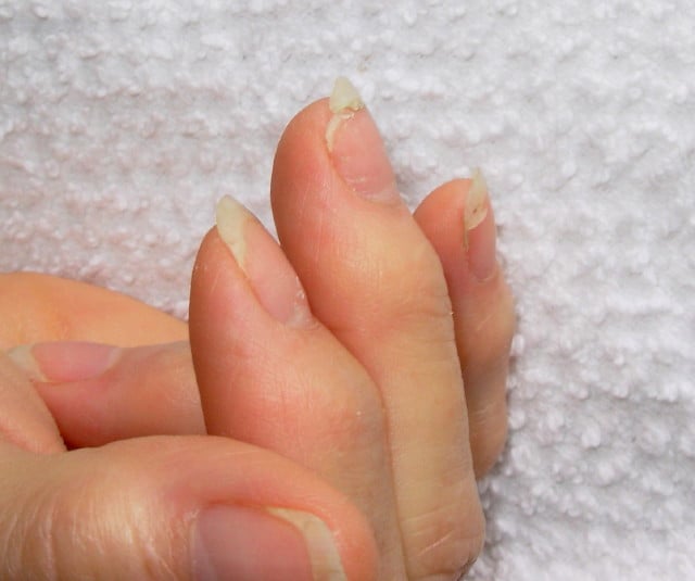
[dropcap]B rittle nails are abnormalities of the nail plate that are characterized by poor nail structure affecting all fingernails and toenails seen as thin, dry nails that easily chip, split, and are difficult to maintain a clean edge. Usually longitudinal ridging occurs from the nail base to the tips.
Q: What is the nail plate?
A: The nail plate is the hard keratin cover protecting the finger tip and toe tip. The nail plate (non-living tissue) is produced by the nail matrix (living tissue) at the base of the nail plate under the lunula (moon), which is the site of brittle nail development.
Nail Anatomy. A. Nail plate; B. lunula; C. root; D. sinus; E. matrix; F. nail bed; G. hyponychium; H. free margin. Courtesy Wikipedia.org
Poor nail structure affecting all nails may be a feature of nutritional deficiency in poor diet or malabsorption such as occurs in celiac disease.
Some other causes are: idiopathic (unknown cause), the result of aging, the effects of certain drugs, or an association with systemic autoimmune disorders such as vitiligo, alopecia areata (with pitting), psoriasis (with pitting), and lichen planus.
External (non-nutritional or disease) causes of dry, brittle nails, such as detergents and cleaners, would only affect fingernails but not toenails.
Note: It has been shown that working with your hands in water or soaking them through activities like swimming does not cause dry, brittle nails but will worsen them.
What Are Brittle Nails In Celiac Disease and/or Gluten Sensitivity?
Beau”s lines in a thin nail. (The tiny brown streaks are splinter hemorrhages due to vitamin C deficiency.)
What Are Horizontal Ridges In Fragile Nails?
Horizontal ridges, also called “beau’s lines,” are abnormalities of the nail plate that appear as rumpling from the base to the tips of nails and are characterized by poor nail structure of both fingernails and toenails.
The nail plate is the hard keratin cover of the finger tip and toe tip which we ordinarily call “nails.” The nail plate is produced by the nail matrix.
Q: Why do Beau’s lines develop in nails?
A: Beau’s lines occur due to temporary cessation of proliferation (growth) of proximal nail matrix at the nail base. As the finger nail grows at the rate of 0.1 mm/day, the time course of the illness can be estimated from the position of the Beau’s line from proximal nail fold.1
Nail Anatomy. .
A. Nail plate; B. lunula; C. root; D. sinus; E. matrix; F. nail bed; G. hyponychium; H. free margin. Courtesy Wikipedia.
Beau’s lines are frequently seen in nutritional deficiency states, bacterial illness, acute stress, and systemic disease. The conditions where Beau’s lines have been described include severe systemic illness, chemotherapy, malnutrition, zinc deficiency, trauma, paronychia, pemphigus, and Kawasaki disease.2 Beau’s lines are commonly seen in patients undergoing chemotherapy.3
This condition of nails was named after Joseph Honoré Simon Beau (1806-1865).
What Are Horizontal Nail Ridges In Celiac Disease and/or Gluten Sensitivity?
Sources:
Naik GS1, Harikrishna J. Beau’s lines. Indian J Med Res. 2013 Jan;137(1):220. [↩]
Naik GS1, Harikrishna J. Beau’s lines. Indian J Med Res. 2013 Jan;137(1):220. [↩]
[dropcap]H ypocalcemia, or low plasma calcium, means the level of calcium in blood is too low to meet metabolic needs of the body for calcium.
Low blood calcium is characterized by bone and tooth demineralization (loss of calcium causing weak teeth and fragile bones), and these impaired functions: nerve conduction, muscle contraction, blood clotting, blood pressure regulation, glycogen to glucose conversion, many hormone actions, many enzyme activities, and acetylcholine production.
Q: Where is calcium found in the body?
A: Calcium is the most abundant mineral in the body, with 99% residing in bones and teeth where it constitutes 40% of skeletal bone weight along with 45% phosphorus. As a component of bone (hard tissue), calcium fulfills a structural role to maintain body size and act as attachments for musculoskeletal tissues. The remaining 1% of calcium is present in blood and soft tissues.
Calcium levels in the blood are maintained within very strict limits by dietary intake, hormonal regulation by the parathyroid gland and a rapidly exchangeable pool in bone tissue.
What Is Hypocalcemia In Celiac Disease and/or Gluten Sensitivity?
Figure on right shows how atherosclerosis impedes blood flow through coronary arteries while blood clots block blood flow. Courtesy Google.
What Is Coronary Artery Disease (CAD)?
[dropcap]C oronary artery disease (CAD), also called ischemic heart disease, is a gradual narrowing of medium and large arteries of the heart by fatty buildups, called atherosclerotic plaques.
It is characterized by slowly developing interference with blood flow to heart tissue itself, resulting in oppressive chest pain called angina and, ultimately, thrombosis (clot) causing heart attack.
The heart is a muscular organ that is working all the time, so it needs a constant supply of oxygen. Oxygen is brought to the working heart tissue by the coronary arteries with each beat of the heart. When heart muscle has to work harder, it needs more oxygen delivered to itself. Lack of oxygen causes pain.
In fact, failure of diseased coronary arteries to deliver adequate oxygen to heart tissue is the most common cause of angina pectoris – substernal pain (under breastbone) or pressure brought on by exertion and relieved by rest.
Thrombosis, or clot formation, occurs when blood cells within a narrowed artery can no longer get through. Trapped, blood cells pile up and block the artery thus triggering a cascade of events called heart attack. Coronary arteries that are narrowed by atherosclerotic plaques can rupture causing injury to the coronary blood vessel resulting in blood clotting which blocks the flow of blood to the heart muscle. Blood clots may form, partially dissolve, and later form again and angina can occur each time a clot blocks blood flow in an artery.1
Q: How does coronary artery disease develop?
A:Coronary artery disease slowly develops from this combination of events:
Dysfunction of epithelial cells that line the inside of arteries cause the vessels to stiffen, and subsequently
Accumulation of lipid (fat) in smooth muscle cells beneath the inside lining of arteries and in foam cells cause buildup of fatty deposits on the inside walls progressing to fibrous plaque formation.
Oxidized low-density lipoprotein (oxLDL), so-called bad cholesterol, and oxysterols play important roles in the development of atherosclerosis. OxLDL triggers the immune system to produce autoantibodies against oxLDL that are detectable in serum. These antibodies are called anti-oxLDL. Anti-oxLDL antibody and oxysterol concentrations are associated with coronary artery stenosis. Oxidative stress may be greatly increased in unstable angina.2 and Chronic inflammation in the general population is a major risk factor for ischemic heart disease.
The pathophysiology of atherosclerosis is, clearly, different in women when compared to the men. The women have a higher risk of blood coagulability making them at high risk for the blood clot formation. In a large number of women endothelial dysfunction, small vessel size and diffuse atherosclerosis have been identified as causes of ischemia without evidence of blockade in the coronary arteries.3
Also, atherosclerotic plaque in women is less fibrotic and contains more lipid filled foam cells, implying greater potential for reversibility but also potentially greater vulnerability for plaque rupture and thrombosis.4
Who is Affected in the General Population?
Coronary artery disease remains the leading cause of death in developed countries despite significant progress in primary prevention and treatment strategies.
It is the leading cause of death in women, as well as an important cause of disability.
Older patients are at particularly high risk of poor outcomes following acute coronary syndrome.5
What Is Coronary Artery Disease In Celiac Disease and/or Gluten Sensitivity?
Ischemic heart disease is the leading cause of death in the United States, making cardiovascular risk assessments and potential interventions or treatments imperative for patients with celiac disease.6
Yasunobu Y, Hayashi K, Shingu T, Yamagata T, Kajiyama G, Kambe M. Coronary atherosclerosis and oxidative stress as reflected by autoantibodies against oxidized low-density lipoprotein and oxysterosis. Atherosclerosis. Apr 2001;155(2):445-53. [↩]
Kunadian V, Ford GA, Bawamia B, Qiu W, Manson JE. Vitamin D deficiency and coronary artery disease: A review of the evidence. Am Heart J. 2014 Mar;167(3):283-291. doi: 10.1016/j.ahj.2013.11.012. Epub 2013 Dec 19. [↩]
Kunadian V, Ford GA, Bawamia B, Qiu W, Manson JE. Vitamin D deficiency and coronary artery disease: A review of the evidence. Am Heart J. 2014 Mar;167(3):283-291. doi: 10.1016/j.ahj.2013.11.012. Epub 2013 Dec 19. [↩]
Kunadian V, Ford GA, Bawamia B, Qiu W, Manson JE. Vitamin D deficiency and coronary artery disease: A review of the evidence. Am Heart J. 2014 Mar;167(3):283-291. doi: 10.1016/j.ahj.2013.11.012. [↩]
Robinson BL, Davis SC, Vess J, Lebel, J. Primary care management of celiac disease. Nurse Practitioner. February 2015: Vol 40 – Issue 2; 28–34. [↩]
[dropcap]U nexpected weight loss is unintentional loss of body mass composition or inability to gain weight marked by decreased serum proteins and increased stool fat.1
What Is Unexpected Weight Loss In Celiac Disease and/or Gluten Sensitivity?
Baby with Allergic Reaction to Peanuts. GFW photo.
What Is Food Allergy?
[dropcap]F ood allergy is an abnormal immune response to food proteins that may damage the small intestinal lining and produce malabsorption of food. The reaction is usually delayed which makes it difficult to identify the offending food that is causing symptoms.
Q: How does food allergy develop?
A: The gastrointestinal tract serves not only to digest and absorb foodstuffs but also to protect the body from unwanted substances. When allergic food substances are eaten, the immune response that is triggered in the gut produces inflammation with symptoms such as pain, vomiting and loose bowels.
Inflammation causes swelling of the gut lining that can interfere with the passage of nutrients through it to the body which results in malabsorption. Malabsorption deprives the body of nutrients needed for normal function.
Symptoms other than digestive may involve skin rashes, hives, and respiratory difficulties that can be distressing and life-threatening.
What Is Food Allergy In Celiac Disease and/or Gluten Sensitivity?