Skip to content

Vitamin C Deficiency

vitamin c gluten celiac disease symptomWhat Is Vitamin C?

[dropcap]V[/dropcap]itamin C, also called ascorbic acid or ascorbate, is an essential water soluble vitamin.

Fresh supplies of vitamin C are required every day to perform vital roles throughout the body among which are the production of connective tissue such as skin, blood vessels and bone, anti-inflammatory responses and anti-oxidant performance. See below for all roles.

Vitamin C fights for us! It has an anti-cancer effect stemming from its role as a potent anti-oxidant in blood plasma and within cells. It also protects nucleic acids (DNA) from oxidative damage and inhibits the formation of nitrosamines (carcinogenic compounds formed in the digestive tract).

Importantly, vitamin C can regenerate vitamin E from an oxidized state after vitamin E performs its own anti-oxidant role against free radicals and vitamin C works together with other anti-oxidants, such as beta-carotene and glutathione, to increase their overall anti-oxidant effect.

A study investigating the specific anti-inflammatory and anti-oxidant micronutrients that reduce oxidative stress found that in adolescents with metabolic syndrome (MetS) the lower the vitamin C level in blood the worse the MetS status and blood uric acid level.  MetS prevalence was estimated at 7% among boys and 3% among girls.1

Vitamin C reverses osteoporosis by reducing or preventing oxidative stress that destroys living bone tissue. Osteoporosis is a disorder of bone inflammation that results in thin, weak bones that may easily fracture. In normal bone structure, vitamin C is required to form collagen, which is living bone tissue.

In humans, vitamin C reduces the duration of common cold symptoms, even if its effect is not clear.2

Vitamin C must be obtained from food daily because our bodies, unlike monkeys and other animals, cannot produce it.

Urinary excretion of vitamin C cannot be detected when vitamin intake is below the required levels. On the other hand, when intake exceeds saturation in the body, the vitamin and/or its metabolites are actively excreted into urine to prevent excessive toxicity of the vitamins.3

What Is Vitamin C Deficiency In Celiac Disease and/or Gluten Sensitivity?

Sources:
  1. TGBeydoun MA, Canas JA, Beydoun HA, Chen X, Shroff MR, Zonderman AB. Serum antioxidant concentrations and metabolic syndrome are associated among U.S. adolescents in recent national surveys. J Nutr. 2012 Sep;142(9):1693-704. doi: 10.3945/jn.112.160416. []
  2. Shaik-Dasthagirisaheb YB, Varvara G, Murmura G, Saggini A, Caraffa A, Antinolfi P, Tete’ S, Tripodi D, Conti F, Cianchetti E, Toniato E, Rosati M, Speranza L,Pantalone A, Saggini R, Tei M, Speziali A, Conti P, Theoharides TC, Pandolfi F. Role of vitamins D, E and C in immunity and inflammation. J Biol Regul Homeost Agents. 2013 Apr-Jun;27(2):291-5. []
  3. Shibata K, Hirose J, Fukuwatari T. Relationship Between Urinary Concentrations of Nine Water-soluble Vitamins and their Vitamin Intakes in Japanese Adult Males. Nutr Metab Insights. 2014 Aug 5;7:61-75. doi: 10.4137/NMI.S17245. eCollection 2014. []

Inflammation

https://glutenfreeworks.s3.amazonaws.com/glutenfreeworks/constipation%20supplements.pdf?X-Amz-Content-Sha256=UNSIGNED-PAYLOAD&X-Amz-Algorithm=AWS4-HMAC-SHA256&X-Amz-Credential=AKIAJE5AFYFVVGZSJR3A%2F20260710%2Fus-east-1%2Fs3%2Faws4_request&X-Amz-Date=20260710T014357Z&X-Amz-SignedHeaders=host&X-Amz-Expires=1000&X-Amz-Signature=6e0f919f337870f072cbf53af97a550f5f44b701b55139621a585c4973b3fd45

inflammation-post-4What Is Inflammation?

[dropcap]I[/dropcap]nflammation is our body’s necessary self-defense response and repair mechanism for these assaults:

1) injuries such as cuts, scrapes, sprains, broken bones, burns, insect bites, toxins; 2) invading organisms such as bacteria; and 3) allergens and food sensitivities such as gluten.

Inflammation can be immediate (acute) or persistent (chronic).

Acute inflammation is marked by increased blood flow, migration of white blood cells, and release of defensive proteins and chemicals to the site of injured tissue. Among these chemicals are free radicals in the immune response to injury that are beneficial yet require the activity of anti-oxidants such as vitamin E and vitamin C to control.

Free radicals are chemical particles containing one or more unpaired electrons, which may be part of the molecule. They cause the molecule to become highly reactive.1

The majority of this response takes place in the first 12 to 24 hours after the assault. The inflammatory process continues until all the damaged tissue or invading germs are removed (usually about 5 days).2

Chronic inflammation is marked by persistence weeks to months or longer after tissue damage. Note: high concentrations of free radicals generated in chronic inflammation may be important causes of damage to cell structures. The defensive activity of anti-oxidants such as vitamin E and vitamin C are required to remove free radicals.

Chronic inflammation increases the risk for systemic diseases such as type II diabetes, obesity, heart disease, high blood pressure, arthritis, osteoporosis, chronic fatigue, migraine, autoimmune disease, and vasculitis which may cause stroke, heart attack or deep vein thrombosis (DVT).

Importantly, chronic inflammation is a risk factor for the onset of cancer.3

Q: Are there blood tests available for detecting inflammation?

A: Yes. Your medical health practitioner can order either or both of the following blood tests that measure the amount of inflammation present although not the source of inflammation. Abnormal is an elevation in blood levels.

  1. C-reactive protein (CRP). This test measure C-reactive proteins that are released into the bloodstream within a few hours of tissue injury or infection. CRPs are cytokines called ‘acute phase reactants,’ meaning first on the scene. The CRP test is also useful to monitor treatment response and flare-ups of chronic inflammatory disease such as vasculitis, systemic lupus, and inflammatory bowel disease.
  2. Erythrocyte sedimentation rate (ESR or sed rate). This test measures the rate of fall of blood cells in a sample tube of blood. An increase in the rate of fall shows inflammation due to an increase of C-reactive proteins in the blood specimen. Alone or with the CRP test, the ESR is especially useful for monitoring inflammation of veins and arteries.

In regards to celiac disease, disappearance of blood antibody levels of tissue transglutaminase IgA (tTG-IgA) indicate that inflammation has also subsided. These antibodies should be checked at 3 months, 6 months if indicated, and one year after diagnosis to monitor healing. On the other hand, raised antibodies indicate that there is definitely ongoing inflammation in the small intestine.

In regards to non-celiac gluten sensitivity, disappearance of blood antibody levels of anti-gliadin IgA and IgG at 3 months, 6 months if indicated, and one year after diagnosis indicate that inflammation has also subsided. On the other hand, raised antibodies indicate that there is definitely ongoing inflammation caused by gluten within the body.

What Is Inflammation In Celiac Disease and/or Gluten Sensitivity?

Sources:
  1. Ruttkay-Nedecky B, Nejdl L , Gumulec J. The Role of Metallothionein in Oxidative Stress. Int. J. Mol. Sci. 2013, 14(3), 6044-6066; doi:10.3390/ijms14036044. []
  2. Taber’s Cyclopedic Medical Dictionary. 19th ed. F A Davis Company. Philadelphia, PA. []
  3. Brighenti E, Giannone FA, Fornari F, Onofrillo C, Govoni M, Montanaro L, Treré D, Derenzini M. Therapeutic dosages of aspirin counteract the IL-6 induced pro-tumorigenic effects by slowing-down the ribosome biogenesis rate. Oncotarget. 2016 Aug 20. doi: 10.18632/oncotarget.11441. []

Systemic Lupus Erythematosus 

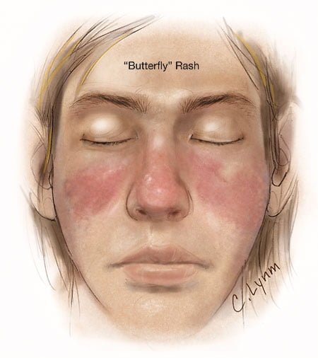
Image showing butterfly rash of SLE. Courtesy JAMA.
Image showing butterfly rash typical of SLE. Courtesy JAMA.

What Is Systemic Lupus Erythematosus?

[dropcap]S[/dropcap]ystemic lupus erythematosus (SLE) is a chronic autoimmune inflammatory disease that is characterized by involvement of multiple organs due to the production of antibodies to components of the cell nucleus.1 SLE has an unpredictable course of acute flare-ups and remissions.

Severity depends on the extent of organs affected with skin and nail involvement, called discoid lupus, being the least serious and inflammmation of the kidney, called lupus nephritis, being the worst.

Nevetheless, a classic presentation is development of a rash over the cheeks and nose that resembles a butterfy with wings spread hence the name “butterfly rash.”

Symptoms are many and varied depending on the tissues affected and are often not specific, for example hair loss has a variety of causes. Symptoms can be confused by co-existence with other autoimmune disease such as Sjogren’s syndrome.

Systemic lupus erythematosus should be managed by a specialist. Symptoms can be controlled with steroid therapy, but this disease can be a cause of premature death  mainly from active disease, organ failure (e.g., kidneys), infection, or cardiovascular disease from accelerated atherosclerosis.

Certain common medicines known to cause drug-induced lupus are:

  • Isoniazid
  • Hydralazine
  • Procainamide

Other less common drugs may also cause the condition. These may include:

  • Anti-seizure medications
  • Capoten
  • Chlorpromazine
  • Etanercept
  • Infliximab
  • Methyldopa
  • Minocycline
  • Penicillamine
  • Quinidine
  • Sulfasalazine

Symptoms tend to occur after taking the drug for at least 3 to 6 months.2

Although there is a strong familial aggregation, the disease is relatively uncommon and most cases are sporadic.1 According to the Center for Diseases (CDC), lupus most commonly affects women of childbearing age but also occurs in infants, children, adolescents, and men with peak occurrence between ages 15 and 40. Blacks (and possibly Hispanics, Asians, and Native Americans) are affected more than Whites.

What Is Systemic Lupus Erythematosus In Celiac Disease and/or Gluten Sensitivity?

Sources:

  1. http://www.cdc.gov/arthritis/basics/lupus.htm [] []
  2. www.nlm.nih.gov/medlineplus/ency/article/000446.htm []

Sjögren’s Syndrome 

Testing the Eyes for Sjogren's Syndrome.
Testing the Eye for Tear Production (L) and Damage to Conjunctiva from Dryness (R).

What Is Sjögren’s Syndrome?

[dropcap]S[/dropcap]jögren’s syndrome is a systemic inflammatory autoimmune disease with a chronic, progressive course that primarily attacks the lacrimal glands of the eye and the salivary glands of the mouth, which are exocrine glands. Exocrine glands secrete the substances they produce through a duct.

Sjögren’s syndrome is ordinarily characterized by dysfunction of the lacrimal glands to produce tears causing dry eye and the salivary glands to produce saliva causing dry mouth, but is not limited by or to these features.

Besides involvement of these exocrine glands, there may be involvement of other parts of the body, termed extraglandular, which may be more severe than eye or mouth features.

There is not yet agreement on classifying Sjögren’s syndrome. Primary and secondary are the two forms generally accepted.1 Both forms can cause mild to severe disease, called the spectrum:

  • Primary Sjögren syndrome. Disease occurs without involvement of other linked autoimmune disorders. In addition to the eyes and mouth, the nose, throat and skin may also be affected and joints, lungs, kidneys, blood vessels, digestive organs and nerves as well.2 Systemic manifestations (other than eyes and mouth) concern a third of patients, including lymphoma in 5% of the patients.3
  • Secondary Sjögren’s syndrome. Disease complicates other autoimmune disease such as systemic lupus erythematosus, rheumatoid arthritis, primary biliary cirrhosis, and celiac disease.

Diagnosis  of Sjögren’s syndrome is made by most doctors based on Schimer’s test for tears and unstimulated whole salivary flow to assess objective eye and oral involvement, since these are the tests most physicians use in clinical practice.4 Specific antibody tests would be  positive for anti-Ro (SSA)/anti-La (SSB) autoantibodies. Sjögren’s syndrome should also be considered when extraglandular manifestations such as vasculitis, polyneuropathy or arthritis occur, even when the patients do not complain of dry eyes and mouth.5

There is no cure for Sjögren’s syndrome. Treatment is aimed to diminish symptoms. For example, steroids and Ibupropen are used to decrease inflammation and pain in joints. Artificial tears and ointments are used for dry eye.

Most people who develop Sjogren’s syndrome are older than 40 years. Nine of ten people with Sjögren’s syndrome are women.2

What Is Sjögren’s Syndrome In Celiac Disease and/or Gluten Sensitivity?

Sources:

  1. Huang YF, Cheng Q, Jiang CM, An S, Xiao L, Gou YC, Yu WJ, Lei L, Chen QM, Wang Y, Wang J. The immune factors involved in the pathogenesis, diagnosis, and treatment of Sjogren’s syndrome. Clin Dev Immunol. 2013;2013:160491. doi: 10.1155/2013/160491. Epub 2013 Jul 9. []
  2. nlm.nih.gov [] []
  3. Fazaa A, Bourcier T, Chatelus E, Sordet C, Theulin A, Sibilia J, Gottenberg JE. Classification criteria and treatment modalities in primary Sjögren’s syndrome. Expert Rev Clin Immunol. 2014 Apr;10(4):543-51. doi: 10.1586/1744666X.2014.897230. []
  4. Cornec D, Saraux A, Cochener B, Pers JO, Jousse-Joulin S, Renaudineau Y, Marhadour T, Devauchelle-Pensec V. Level of agreement between 2002 American-European Consensus Group and 2012 American College of Rheumatology classification criteria for Sjogren’s syndrome and reasons for discrepancies. Arthritis Res Ther. 2014 Mar 19;16(2):R74. []
  5. Witte T. Pathogenesis and diagnosis of Sjögren’s syndrome. Z  Rheumatol. 2010 Feb;69(1):50-6. doi: 10.1007/s00393-009-0519-2. []

Polymyositis

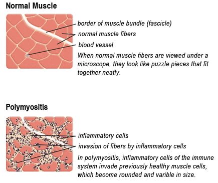
Drawing of Biopsy Showing Muscle Fibers Invaded by Immune Cells. Courtesy MDA.org

What Is Polymyositis?

[dropcap]P[/dropcap]olymyositis is a body-wide connective tissue disease resulting from autoimmune attack of skeletal muscles that is characterized by inflammatory and degeneratory changes. The course is unpredictable being marked by spontaneous flare-ups and remissions.

Polymyositis can begin slowly or abruptly according to the factor that is triggering the onset such as infection, medications like phenytoin, and autoimmune disease.

Progressive muscle weakness starts in the proximal skeletal muscles (muscles closest to the trunk of the body).

Skeletal muscles, also called voluntary, are muscles that move the body as we want, such as walking and  lifting objects, as opposed to those we cannot voluntarily control, such as the muscles of digestion. 

Q: What are the degeneratory changes in skeletal muscles?

A: In polymyositis, degeneratory changes in skeletal muscles means that muscles are being destroyed (called necrosis), resulting in fibrosis, or scarring. When scar tissue takes the place of lost muscle tissue, it cannot act like muscle to contract and relax.   Muscle destruction is what causes muscle pain and weakness.

After the clinical work-up of exams and blood studies to determine muscle damage, the diagnosis of polymyositis is confirmed by muscle biopsy. See image at above left. The black dots are inflammatory cells. Edema (fluid) between cells caused by inflammation pushes muscle fibers apart.

There is no cure for polymyositis, but the symptoms can be treated.  Options include medication, physical therapy, exercise, heat therapy (including microwave and ultrasound), orthotics and assistive devices, and rest.  The standard treatment for polymyositis is a corticosteroid drug, given either in pill form or intravenously.  Immunosuppressant drugs, such as azathioprine and methotrexate, may reduce inflammation in people who do not respond well to prednisone. 

Periodic treatment using intravenous immunoglobulin can also improve recovery.  Other immunosuppressive agents used to treat the inflammation associated with polymyositis include cyclosporine A, cyclophosphamide, and tacrolimus.  Physical therapy is usually recommended to prevent muscle atrophy and to regain muscle strength and range of motion.1

Diagnosis is based on elevated muscles enzymes, increased urinary creatine level, and electromyograph abnormalities.

Polymyositis can affect people at any age. It is most common in adults between ages 50 and 70, and in children ages 5 to 15. It affects women twice as often as men and is more common in African Americans than Caucasians.2 The major causes of death from polymyositis are cancer and lung disease, including pneumonia.  The 5-year mortality rate can be as high as 1 in 5 patients.2

What Is Polymyositis In Celiac Disease and/or Gluten Sensitivity?

Sources:

  1. National Institute of Neurological Disorders and Stroke []
  2. http://www.nlm.nih.gov/medlineplus/ency/article/000428.htm [] []

Cystic Fibrosis 

Bronchiectasis in Cystic Fibrosis. Courtesy Dr Jerry Nick National Jewish Medical and Research Center, Denver
Lung Tissue in Bronchiectasis.  Courtesy Dr Jerry Nick National Jewish Medical and Research Center, Denver

What Is Cystic Fibrosis?

[dropcap]C[/dropcap]ystic fibrosis is a genetic disease of the exocrine glands which causes the body to produce abnormally thick and sticky mucus and results in progressive damage to the respiratory system and digestive problems.

Severity of disease depends on the particular genetic mutation of affected individuals.

Q: How does the abnormal mucus damage the lungs and digestive tract?

A: In all cases of cystic fibrosis, sticky mucus build-up in the bronchioles (small breathing passages) of the lungs causes COPD (chronic obstructive pulmonary disease) that results in life-threatening lung infections. 

In the pancreas, the abnormal mucus damages its ability to produce required enzymes to digest food, causing failure to adequately digest fats, carbohydrates and protein which are vital for life.

Other tissues that may be involved are sweat glands, sinuses, and the male reproductive system, causing infertility.

Millions of Americans carry the defective cystic fibrosis gene, but do not have any symptoms. That is because cystic fibrosis requires the inheritance of two defective cystic fibrosis genes — one from each parent who carries it. An estimated 1 in 29 Caucasian Americans have the cystic fibrosis gene.

The disease is the most common, deadly, inherited disorder affecting Caucasians in the United States. It is more common among those of Northern or Central European descent. About 30,000 children and adults in the United States have cystic fibrosis while more than 10 million people carry the gene.1

There is no cure yet for cystic fibrosis, however, treatments have greatly improved in recent years. The goals of treatment include:

  • Preventing and controlling lung infections.
  • Loosening and removing thick, sticky mucus from the lungs.
  • Preventing or treating blockages in the intestines.
  • Providing enough nutrition.
  • Preventing dehydration (a lack of fluid in the body).2

What Is Cystic Fibrosis In Celiac Disease and/or Gluten Sensitivity?

Sources:

  1. www.ncbi.nlm.nih.gov/pubmedhealth/PMH0001167/ []
  2. National Heart, Blood, and Lung Institute []

Hemochromatosis (Iron Overload Disease)

Hemochromatosis
Microscopic View of Hemochromatosis Stained Blue. Courtesy Wikimedia

What Is Hemochromatosis?

[dropcap]H[/dropcap]emochromatosis, also called iron overload liver disease, is a common inherited disease in the Caucasian population that is characterized by increased iron deposition within the tissues (overload) associated with injury to them.

Hemochromatosis is an autosomal recessive disease, meaning a pair of abnormal genes are inherited from each parent. Parents are unaffected because they carry only one gene.

Q: Where is iron deposited?

A: In hemochromatosis, more iron is absorbed from the small intestine than is needed by the body. Subsequently, because the body has no satisfactory means to release iron overload, excess iron is deposited in various organs such as the liver causing cirrhosis, joints causing arthritis, and the pancreas causing diabetes mellitus.1

Consumption of alcoholic drinks with food should be avoided because alcohol increases leaky gut and greatly increases the absorption of iron. On the flip side, eggs and foods containing calcium such as milk and cheese are beneficial for hemochromatosis because they impair the absorption of iron. Another iron inhibitor is food that contains phytic acid which includes the bran and outer layer of grains, seeds, nuts, peas, beans, and lentils.

Iron overloading, as measured by a random (non-fasting) elevated transferrin saturation value, is estimated to occur in 1 to 6 people per 100 in the United States according to the CDC (Center for Disease Control and Prevention). Too much iron increases the risk for metabolic syndrome, type 2 diabetes mellitus, cancer, liver disease, and osteoporosis.

Medical treatment to remove excess iron from the body uses phlebotomy that draws blood through a vein and/or certain injectable drugs (deferoxamine and deferairox) that bind to iron.

What Is Hemochromatosis In Celiac Disease and/or Gluten Sensitivity?

Sources:

  1. www.irondisorders.org []