Skip to content

Dementia

DementiaWhat Is Dementia?

Dementia is the term used to describe a group of symptoms that show significant deterioration of an individual’s intellectual and social abilities.

The deterioration in intellectual function is progressive and is characterized by memory and cognitive impairment involving deficits in reasoning, judgment, abstract thought, comprehension, learning, use of language, and task execution.

Some types of dementia can be reversed,  while most types of dementia are degenerative or nonreversible.

Q: What causes dementia?

A: There are  many differing causes of dementia.  Here are some causes according to nonreversible and reversible:

  • Nonreversible dementia may not be turned back due to these conditions:
  • Alzheimer’s disease is the most common type of degenerative dementia caused by abnormal protein structures in certain areas of the brain. 
  • Lewy body disease is a leading cause of dementia in elderly adults.
  • Vascular dementia due to many small strokes.
  • Medical conditions: Huntington’s disease, multiple sclerosis, infections that can affect the brain, such as HIV/AIDS and Lyme disease, Parkinson’s disease, Pick’s disease, and progressive supranuclear palsy.
  • Reversible dementia may be stopped or reversed if these conditions are found soon enough:
  • Brain injury.
  • Brain tumors.
  • Chronic alcohol abuse.
  • Changes in blood sugar, sodium, and calcium levels.
  • Changes that can occur with celiac disease, diabetes, thyroid disease, and other metabolic disorders.
  • Nutritional deficiencies.
  • Use of certain medications, including cimetadine and some cholesterol-lowering medications.1

What Is Dementia In Celiac Disease and/or Gluten Sensitivity?

Sources:
  1. http://www.ncbi.nlm.nih.gov/pubmedhealth/PMH0001748/ []

Helicobacter Pylori Infection (H. Pylori)

What Is Helicobacter Pylori (H. Pylori) Infection? Helicobacter pylori infection is a potentially deadly stomach disease characterized by chronic superficial inflammation and ulcerations in 100% of infected patients. This infection disrupts normal defense and repair… 

Gastric (Stomach) Ulcer

Photo by gastroscopy showing ulcer in the antrum area of the stomach.
Photo by gastroscopy showing ulcer in the antrum area of the stomach (lower area).

What Is A Gastric Ulcer?

Gastric ulcer is a painful stomach disorder characterized by an open sore involving the mucosa lining and deeper muscle layer of the stomach.

Gastric ulcer is associated with lymphocytic gastritis which is inflammation of the mucosal lining of the stomach. The thick mucosal lining normally protects the stomach from the erosive action of stomach acid.

Q: How do ulcers develop?

A: Ulcers develop if  hydrochloric acid secreted by the gastric glands of the stomach for the purpose of digesting food damages the normally resistant mucosal walls of the stomach. In the reverse, ulcers may be accompanied by achlorhydria (insufficient acid production).

Damage occurs when there is a predisposing factor that alters the health of the mucosal lining. The most common cause is infection with a bacteria called h. pylori bacter, stress and chronic use of the pain relievers aspirin and non-steroidal drugs like ibuprofen.

Smoking tocacco and consuming alcohol aggravate an ulcer but do not cause it to develop.

The most common location for ulcer formation is along the stomach antrum which is the area of the stomach before the pylorus, the lower region that empties liquid stomach contents into the small intestine.

What Is A Gastric Ulcer In Celiac Disease and/or Gluten Sensitivity?

Vitamin B3 (Niacin) Deficiency

SumptuousTuna For Niacin.
Sumptuous Tuna For Niacin.

What Is Vitamin B3 (Niacin)?

Niacin is an essential water-soluble B vitamin that is required by all cells of the body.

During digestion of food containing it, niacin (the form in food) is changed in the small intestines to the active form niacinamide (niacin plus an amide group), which is then absorbed into the bloodstream. 

Niacinamide is converted by the body into co-enzymes which are present in all cells. These are niacinimide adenine dinucleotide (NAD) and NADP. NADP is formed when the body adds a phosphate to NAD.

Q: How do these enzymes work?

A: These enzymes function in oxidation-reduction reactions essential for release of energy from carbohydrates, fats, and proteins and are needed as components for more than 200 enzymes involved in metabolism.

In addition to producing energy, niacinamide is essential for healthy skin and the mucosal lining of the digestive tract, normal functioning of the brain and nervous system, and production of steroid hormones in adrenal glands and hormones in sex glands.  Functions are more fully described below.

Urinary excretion of niacin cannot be detected when vitamin intake is below the required levels. On the other hand, when intake exceeds saturation in the body, the vitamin and/or its metabolites are actively excreted into urine to prevent excessive toxicity of the vitamins.1

What Is Niacin Deficiency In Celiac Disease and/or Gluten Sensitivity?

Sources:
  1. Shibata K, Hirose J, Fukuwatari T. Relationship Between Urinary Concentrations of Nine Water-soluble Vitamins and their Vitamin Intakes in Japanese Adult Males. Nutr Metab Insights. 2014 Aug 5;7:61-75. doi: 10.4137/NMI.S17245. eCollection 2014. []

Peripheral Neuropathy 

Microscopic Slide of Lymphocytic Colitis. Courtesy Quizlet.com
Microscopic  Slide of Biopsy Sample Showing Lymphocytic Colitis. Courtesy Quizlet.com

What Is Lymphocytic Colitis?

L ymphocytic colitis is a microscopic inflammation of the large intestinal mucosa with infiltration of lymphocytes (IELs)  that is characterized by non-bloody secretory diarrhea.

Secretory diarrhea describes bowel movements that consist of a large volume of liquid stool.

Q: What are IELs?

A: IELs is an abbreviation for intraepithelial lymphocytes, which are white blood cells that infiltrate within epithelial cells or between them. Epithelial cells form the surface mucosa of the large intestine also called the colon.

The histopathological criteria (biopsy) for lymphocytic colitis are a density of at least 20 IELs per 100 surface epithelial cells; chronic inflammatory infiltrate of mononuclear cells in the lamina propria; epithelial damage; and a subepithelial collagen layer of less than 10 µm. The increased collagen band consists basically of collagen type I and III, which are the subtypes produced by repair functions, indicating a reactive origin.1That is, the mucosa is reacting to some irritative substance.

Up to 10% of adults undergoing colonoscopy for investigation of chronic diarrhea and having visibily normal appearing mucosa may have lymphocytic colitis.2

Bile acid malabsorption has been shown to coexist in 60% of patients with lymphocytic colitis.1

Lymphocytic colitis (LC) is categorized as primary or secondary.  Primary LC is a clinical and histopathological disease of unknown cause. Secondary LC may develop as the result of iritating factors acting on the colon such as smoking or many medications.  In one study, the most common drug treatments as a percentage of the study group were corticosteroids (32.1%), proton pump inhibitors (26.0%), antidepressant drugs, specifically selective serotonin reuptake inhibitors (21.4%), angiotensin-converting enzyme inhibitors or angiotensin II receptor antagonists (18.3%), statins (17.6%), thyroid hormones (17.6%), and beta-blockers (16.0%).3

Secondary lymphocytic colitis is associated with several concomitant diseases including celiac disease. This is why lymphocytic changes must be interpreted with caution before considering them as a separate entity of autoimmune origin, instead of secondary reactions to ischemia and toxic stimulants. Efforts must be made to better classify and diagnose patients with real, primary lymphocytic colitis to avoid over-prescription of corticosteroids for treatment.3

What Is Lymphocytic Colitis In Celiac Disease and/or Gluten Sensitivity?

Sources:
  1. Ohlsson B. New insights and challenges in microscopic colitis. Therap Adv Gastroenterol. 2015 Jan;8(1):37-47. doi: 10.1177/1756283X14550134. [] []
  2. Abdo AA, Urbanski SJ, Beck PL. Lymphotcytic and collagenous colitis: the emerging entity of microscopic colitis. An update on pathophysiology, diagnosis and management. Canadian Journal of Gastroenterology. Jul 2003;17(7):425-32. []
  3. Roth B, Manjer J, Ohlsson B. Drug Target Insights. 2013 Aug 11;7:19-25. doi: 10.4137/DTI.S12109. [] []

Autism and Learning Disabilities

Malignant lymphoma high grade B-cell. Courtesy Wikimedia

What Is B-Cell Non-Hodgkin’s Lymphoma?

B cell non-Hodgkin’s lymphoma is a malignant, monoclonal (arising from a single cell) proliferation of lymphocytes that is preceded by lymphadenopathy and characterized by varying, less predictable spread than Hodgkin’s disease.

Lymphadenopathy is enlargement of lymph nodes greater than 1.5 cm caused by activation and increased production of lymphocytes and phagocytes (type of white blood cell that engulfs pathogens during infection) or invasion by a tumor.

Q: How does this type of lymphoma develop?

A: 80% to 85% of non-Hodgkin’s lymphoma arise from B-lymphocytes (B-cells).

What Is B Cell Non-Hodgkin’s Lymphoma In Celiac Disease and/or Gluten Sensitivity?

Apathy

Cows Milk enteropathyWhat Is Milk Intolerance (Bovine Beta Casein Enteropathy)?

B ovine beta casein enteropathy is characterized by raised serum IgA antibodies to bovine beta casein found by blood testing and damage to mucosa of the jejunum found by biopsy.

The jejunum is the second section of the small intestine, between the duodenum and the ileum.

Mucosa damage is similar to damage caused by gluten.

Q: What is bovine beta casein?

A: Bovine beta casein is a protein found in cow’s milk.

An immune reaction to protein in milk is totally different from lactose intolerance. Lactose intolerance develops from deficiency of an intestinal enzyme called lactase which is required to digest lactose, the sugar in milk.

What Is Milk Intolerance In Celiac Disease and/or Gluten Sensitivity?

Muscle Pain and Tenderness, Chronic 

Reproduction of a lithograph plate showing inside of the stomach from Gray's Anatomy. Courtesy Wikipedia Commons.
Reproduction of a lithograph plate showing inside of the stomach from Gray’s Anatomy. Courtesy Wikipedia Commons.

What Is Delayed Gastric Emptying?

D elayed gastric emptying is a stomach motility or movement disorder characterized by abnormally slow movement of gastric contents from the stomach through the pyloric sphincter into the duodenum, causing dyspepsia.

Q: What determines how fast the stomach empties?

A: How fast the stomach empties depends on the pressure of strong coordinated muscle contractions in the top region of the stomach propelling chyme against resistance at the pylorus (base region of the stomach).

Chyme is food that has been dissolved and thoroughly mixed with stomach secretions.

In the digestion of carbohydrate, protein and fat, protein leaves the stomach first (1 hour), then carbohydrates (1 1/2 to 2 hours), and fat takes longest to digest (2-4 hours). Plain water is able to pass through the pylorus within 5 minutes.

What Is Delayed Gastric Emptying In Celiac Disease and/or Gluten Sensitivity?

Adenocarcinoma Of Small Intestine (Cancer)

Section of small bowel surgically removed for adenocarcinoma that grew through the wall. By: CDC/ Dr. Edwin P. Ewing, Jr.

What Is Adenocarcinoma Of Small Intestine?

Adenocarcinomas are malignant tumors, or cancer, of the small bowel arising out of glandular tissue. They fall in the category of rare neoplasm, comprising only 3% of all gastrointestinal malignancies.

Primary adenocarcinoma is the most common histological (cell) subtype constituting 35–50% of cases.1 

Q: What does adenocarcinoma look like?

A: Adenocarcinoma may manifest as strictures, nodules, excavating masses, or annular lesions.2

What Is Adenocarcinoma Of Small Intestine In Celiac Disease and/or Gluten Sensitivity?

Sources:
  1. Benhammane H, El M’rabet FZ, Serhouchni KI, El yousfi M, Charif I, Toughray I, et al. Small Bowel Adenocarcinoma Complicating Coeliac Disease: A Report of Three Cases and the Literature Review. Case Rep Oncol Med. 2012; 2012: 935183. Published online 2012 December 1. doi: 10.1155/2012/935183 []
  2. Ramachandran I, Sinha R, Rajesh A, Verma R. Multidetector row CT of small bowel tumors.  Clinical Radiology. 2007; 62:607-614. []

Tongue – Fiery Red, Smooth, Burning

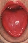
niacin deficiencyWhat Is A Fiery Red, Smooth, Burning Tongue?

A fiery red, smooth, burning tongue is an alteration in tongue tissue that is characteristic of advanced niacin deficiency.1

Q: How does niacin deficiency cause the tongue to be red and sore?

A: Niacin is an essential B vitamin that is required for a healthy tongue and by all body cells as well. Deficiency first shows in tissues with rapid cellular turnover, such as mucosal cells of the tongue.

When absorbed from the small intestines, niacin (the form in food) becomes the active form niacinamide. Niacinamide is converted by the body into co-enzymes which are present in all cells. These enzymes function in oxidation-reduction reactions essential for release of energy from carbohydrates, fats, and proteins and are needed as components for more than 200 enzymes involved in metabolism.

In addition to producing energy, niacinamide is essential for healthy skin and the mucosal lining of the digestive tract, normal functioning of the brain and nervous system, and production of steroid hormones from adrenal glands and hormones from sex glands.

What Is A Fiery Red, Smooth, Burning Tongue In Celiac Disease and/or Gluten Sensitivity?

Sources:
  1. Krause’s Food, Nutrition, & Diet Therapy. 10th Edition. Kathleen Mahan, Sylvia Escott-Stump. 2000. W.B. Saunders Company. []