What Is Vitiligo? V itiligo is a pigmentation disorder of the skin characterized by permanent loss of melanocytes in defined areas and, in some patients, antibodies to melanin. Vitiligo has significant psychological impact if occurring before…
Mucosa in refractory celiac disease immunostained sequentially for CD3 (alkaline phosphatase-blue) and CD8 (peroxidase-brown). Most intraepithelial lymphocytes are CD3+, CD8-. Courtesy pubcan.org
What Is Refractory Celiac Disease?
R efractory celiac disease, formerly called refractory sprue, is a severe complication characterized by persistence of symptoms and intestinal inflammation despite gluten free diet after 12 months.1
Refractory celiac disease appears in two forms, ulcerative jejunitis (RCD I) and cryptic intestinal T-cell lymphoma (RCD II).
Patients with RCD I seem to profit from immunosuppressive treatment, but positive response to corticosteroid treatment does not exclude underlying enteropathy–associated T-cell lymphoma (EATL).
Patients with RCD II have a high percentage of abberant T-cells and is usually resistant to medical therapies. The presence of an aberrant clonal intraepithelial T-cell population has led to the designation of refractory celiac disease with this population as a cryptic intestinal T-cell lymphoma, characterized by frequent dissemination to the blood and the entire gastrointestinal lining.2
Refractory sprue may occur after an initial response to gluten free diet or without any evidence of preexisting celiac disease. All other causes of malabsorption must be excluded, such as collagenous colitis.
In a subgroup of patients with enteropathy-associated T-cell lymphoma (EATL) there is progressive deterioration of a refractory form of celiac disease. The prognosis is poor, although some patients respond to corticosteroids and immunosuppressive agents.3
A nationwide Finnish study showed that patients of male gender, older age, severe symptoms or seronegativity (negative antibody result) at the diagnosis of celiac disease are at risk of future refractory coeliac disease and should be followed up carefully.4
Chorea has been described as a paraneoplastic phenomenon in patients with non-Hodgkin’s lymphoma and has been described as associated with lymphoma arising from a background of refractory celiac disease. The finding of chorea in association with celiac disease should prompt a search for possible underlying intestinal T-cell lymphoma.5
How Prevalent Is Refractory Celiac Disease?
Sources:
Murray JA, The widening spectrum of celiac disease. American Journal of Clinical Nutrition. Mar 1999;69 (3):354-365. [↩]
Culliford AN, Green PH. Refractory sprue. Current Gastroenterology Reports. Oct 2003;5(5):373-8. [↩]
Culliford AN, Green PH. Refractory sprue. Current Gastroenterology Reports. Oct 2003;5(5):373-8. [↩]
Ilus T, Kaukinen K, Virta LJ, Huhtala H, Mäki M, Kurppa K, Heikkinen M, Heikura M, Hirsi E, Jantunen K, Moilanen V, Nielsen C, Puhto M, Pölkki H, Vihriälä I, Collin P. Refractory coeliac disease in a country with a high prevalence of clinically-diagnosed coeliac disease. Aliment Pharmacol Ther. 2014 Feb;39(4):418-25. doi: 10.1111/apt.12606. [↩]
Kitiyakara T, Jackson M, Gorard DA. Refractory coeliac disease, small-bowel lymphoma and chorea. J R Soc Med. 2002 Mar;95(3):133-4. [↩]
CT Scan of Parathyroid Carcinoma Displacing the Trachea by Doina Piciu. Courtesy Orphanet Journal of Rare Diseases
What Is Parathyroid Carcinoma?
P arathyroid carcinoma is a slow growing rare malignancy involving overactive parathyroid glands and is characterized by profound hypercalcemia (elevated blood calcium level), parathyroid hormone levels of more than 3 times upper normal limits, and palpable neck mass.1
Parathyroid carcinoma is a cause of severe hyperparathyroidism. Hyperparathyroidism results from overproduction of parathyroid hormone by the parathyroid glands.
Research comparing patients with carcinoma to patients with primary hyperparathyroidism showed that patients with parathyroid carcinoma had significantly higher serum parathyroid hormone and calcium levels compared with patients with primary hyperparathyroidism.2
Q: Why is the blood calcium level elevated?
A: Blood calcium level is elevated due to overproduction of parathyroid hormone by the diseased gland(s).
The function of parathyroid hormone (PTH) is to keep calcium blood levels normal, which it does by drawing calcium out of bones as needed. The problem of too much PTH is that bone is constantly robbed of calcium and this depletion causes bone demineralization, resulting in osteopenia progressing to osteoporosis. Of course, weakened bones are subject to breakage with light trauma.
On the other hand, too much calcium in the blood can cause kidney stone formation because urine production is the way the body excretes excess calcium. That is, highly concentrated calcium precipitates out of the urine solution to form stones.
With an estimated incidence of 0.015 per 100,000 population and an estimated prevalence of .005% in the United States, parathyroid cancer is one of the rarest of all human cancers according to the National Cancer Institute.
What Is Parathyroid Carcinoma In Celiac Disease and/or Gluten Sensitivity?
Sources:
Schaapveld M, Jorna FH, Aben KK, Haak HR, Plukker JT, Links TP. Incidence and prognosis of parathyroid gland carcinoma: a population-based study in The Netherlandsestimating the preoperative diagnosis. Am J Surg. 2011 Nov;202(5):590-7. doi: 10.1016/j.amjsurg.2010.09.025. Epub 2011 Aug 20. [↩]
Schaapveld M, Jorna FH, Aben KK, Haak HR, Plukker JT, Links TP. Incidence and prognosis of parathyroid gland carcinoma: a population-based study in The Netherlandsestimating the preoperative diagnosis. Am J Surg. 2011 Nov;202(5):590-7. doi: 10.1016/j.amjsurg.2010.09.025. Epub 2011 Aug 20. [↩]
Swollen Tongue Causing Tooth Indentations. Notice the Accompanying Denuded Area Due to Riboflavin Deficiency and Mild Candida Overgrowth. GFW
What Is A Pale, Smooth, Burning Tongue?
A pale, smooth, burning tongue is an alteration in tongue tissue characteristic of iron deficiency. The tongue is also swollen.1
Additionally, the sore tongue surface may be invaded by candida yeast which takes advantage of the sore tissue.
Iron deficiency itself increases susceptibility to infection.
Q: What is iron deficiency?
A: Iron deficiency results when the level within cells is too low to meet metabolic needs of the body for this mineral.
Deficiency is characterized by impaired red blood cell formation, free-radical disposal, oxygenation of cells, immune response to infection, enzyme activity, cognitive performance, digestion, nail structure, and fetal health.2
Iron is an essential mineral that is required for normal body function. Almost two-thirds of iron in the body is found in hemoglobin, the protein in red blood cells that carries oxygen to tissues. Smaller amounts of iron are found in myoglobin, a protein that helps supply oxygen to muscle, and in enzymes that assist biochemical reactions.
Iron is also found in proteins that store iron for future needs and that transport iron in blood. Iron stores are regulated by intestinal iron absorption.3
What Is A Pale, Smooth, Burning Tongue In Celiac Disease and/or Gluten Sensitivity?
Microscopic Image Showing a Pink Collagen Band in Collagenous Colitis.
What Is Collagenous Colitis?
C ollagenous colitis is a disease of the large intestine (colon) that is characterized by microscopic inflammation of the surface mucosal lining and an abnormally thickened collagen band of tissue that develops wthin the lining of the colon.
The thicker than normal layer of collagen of at least 10 µm (reference value: 2–7 µm) can vary in different locations. Inflammation occurs with increased numbers of lymphocytes (white blood cells) and plasma cells and epithelial (surface cell) damage. These changes can only be seen under microscopic examination of multiple biopsied tissue samples taken during a colonoscopy procedure.
Q: What is collagen?
A:Collagen is a strong, fibrous protein found in connective tissue of the colon and many other tissues such as tendons. The normal basement membrane in the bowel consists mainly of collagen type IV, laminin, and fibronectin. The increased collagen band observed in collagenous colitis consists basically of collagen type I and III, which are the subtypes produced by repair functions, indicating a reactive origin to some irritant or drug.1
The biopsies should preferably be taken from the ascending colon, since the pathological hallmarks may be absent in the descending colon, and in the normally occurring thicker collagen layer in the rectosigmoid region.1 Inflammation of the ileum (last segment of the small intestine next to colon) is common.2
Endoscopy and radiological (x-ray) examinations are usually normal.3
Autoimmune disorders are frequently seen in adult patients with collagenous colitis.4 In the study below by Koskela et al. concomittent autoimmune diseases were present in 53% of patients with collagenous colitis.5
Importantly, the finding of collagenous colitis in patients with autoimmune diseases may reflect the treatment with NSAIDs (non-steroidal anti-inflammatory drugs), such as Ibuprofin and aspirin, PPIs (proton pump inhibitors), and other drugs. However, if secondary forms of collagenous colitis are not taken into consideration, underlying, treatable diseases may be overlooked, while only the gastrointestinal symptoms are treated symptomatically or with budesonide (a steroid).6
Treatment with budesonide steroid is efficacious irrespective of bile acid malabsorption.7
Budesonide at a mean dose of 4.5 mg/day maintained clinical remission for at least 1 year in the majority of patients with collagenous colitis and preserved health-related quality of life without safety concerns. Treatment extension with low-dose budesonide beyond 1 year may be beneficial given the high relapse rate after budesonide discontinuation.8
See below for nutritional deficiency problems caused by steroid usage and steps to be taken for correction.
What Is Collagenous Colitis In Celiac Disease and/or Gluten Sensitivity?
Sources:
Ohlsson B. New insights and challenges in microscopic colitis. Therap Adv Gastroenterol. 2015 Jan;8(1):37-47. doi: 10.1177/1756283X14550134. [↩] [↩]
Bjørnbak C, Engel PJ, Nielsen PL, Munck LK. Microscopic colitis: clinical findings, topography and persistence of histopathological subgroups. Aliment Pharmacol Ther. 2011 Nov;34(10):1225-34. doi: 10.1111/j.1365-2036.2011.04865.x. [↩]
Abdo AA, Urbanski SJ, Beck PL. Lymphotcytic and collagenous colitis: the emerging entity of microscopic colitis. An update on pathophysiology, diagnosis and management. Canadian Journal of Gastroenterology. Jul 2003;17(7):425-32. [↩]
Leung ST, Chandan VS, Murray JA, Wu TT. Collagenous gastritis: histopathologic features and association with other gastrointestinal diseases. Am J Surg Pathol. 2009 May;33(5):788-98. doi: 10.1097/PAS.0b013e318196a67f. [↩]
Koskela RM, Niemela SE, Karttunen TJ, Lehtola JK. Clinical characteristics of collagenous and lymphocytic colitis. Scandanavian Journal of Gastroenterology. Sep 2004;39(9):837-45. [↩]
Ohlsson B. New insights and challenges in microscopic colitis. Therap Adv Gastroenterol. 2015 Jan;8(1):37-47. doi: 10.1177/1756283X14550134. [↩]
Bjørnbak C, Engel PJ, Nielsen PL, Munck LK. Microscopic colitis: clinical findings, topography and persistence of histopathological subgroups. Aliment Pharmacol Ther. 2011 Nov;34(10):1225-34. doi: 10.1111/j.1365-2036.2011.04865.x. [↩]
Münch A, Bohr J, Miehlke S, et al. Low-dose budesonide for maintenance of clinical remission in collagenous colitis: a randomised, placebo-controlled, 12-month trial. Gut. 2014 Nov 25. pii: gutjnl-2014-308363. doi: 10.1136/gutjnl-2014-308363. [↩]
H ypertransaminasemia is a chronic condition of elevated blood liver transaminase enzymes, commonly called “liver enzymes,” that signifies hepatocellular (liver) injury.
Q: What are serum transaminases?
A: Transaminases are the liver enzymes ALT and AST. ALT is the abbreviation for alanine aminotransferase enzyme and AST is the abbreviation for aspartate aminotransferase enzyme.
Transaminases are commonly measured in routine blood tests to determine liver function. Elevated ALT and AST transaminases indicate inflammation of the liver. Other specific tests must follow to determine the cause of inflammation.
What Is Hypertransaminasemia In Celiac Disease and/or Gluten Sensitivity?
Angina pectoris, or simply angina, is a coronary syndrome characterized by an oppressive substernal pain (pain under breastbone) or pressure brought on by exertion and relieved by rest that results from failure of coronary arteries to deliver adequate oxygen to heart tissue due to ischemic heart disease.
Q: Why do coronary arteries fail to deliver adequate oxygen to heart tissue?
A: Coronary arteries are the blood vessels that serve the heart. In angina, these vessels fail to deliver adequate oxygen to heart tissue because they are narrowed or blocked by fatty buildups, called atherosclerotic plaques or by a blood clot which impair their ability to carry adequate blood that carries the oxygen. Diseased coronary arteries cannot deliver adequate oxygenated blood pumped by the heart to its own muscle cells.
The heart is a muscular organ that is working all the time without rest, so it needs a constant supply of oxygen. When heart muscle has to work harder, it needs more oxygen. Lack of oxygen causes pain which makes the affected person stop activity and rest.
Angina can be stable or unstable. Unstable angina is much more serious and can be life-threatening.
Stable angina produces predictable pain and responds to rest and/or medication. It is less serious than unstable angina but can be very painful or uncomfortable. Anything that makes the heart muscle need more oxygen can cause an angina attack in someone with heart disease, including: smoking, cold weather, exercise, emotional stress, obesity, and large meals. Other causes of angina include: abnormal heart rhythms (usually ones that cause the heart to beat quickly), anemia, coronary artery spasm, heart failure, heart valve disease, and hyperthyroidism (overactive thyroid).1
Unstable angina produces unpredictable pain that may occur at rest, lasting more than 20 minutes. It is more severe than stable angina and less responsive to medication. Atherosclerosis is by far the most common cause of unstable angina. Oxidized low-density lipoprotein, so-called bad cholesterol, and oxysterols play an important role in atherogenesis, the development of atherosclerosis. Coronary arteries that are narrowed by atherosclerotic plaques can rupture causing injury to the coronary blood vessel resulting in blood clotting which blocks the flow of blood to the heart muscle. Blood clots may form, partially dissolve, and later form again and angina can occur each time a clot blocks blood flow in an artery. People with unstable angina are at increased risk of having a heart attack.2
What Is Angina In Celiac Disease and/or Gluten Sensitivity?
Elevated bone alkaline phosphatase (BALP) is a laboratory result that indicates an abnormal blood level of this bone enzyme.
A bone alkaline phosphatase blood level is one of the most frequently used biochemical markers of bone formation.
Q: Why is the purpose of bone alkaline phosphatase?
A: Bone alkaline phosphatase is produced by bone cells called osteoclasts in normal bone maintenance for the purpose of breaking down old or damaged bone so that other bone cells called osteoblasts can fill in the excavated areas with new bone. This process keeps bone stong and healthy.
Elevated bone alkaline phosphatase shows that more bone is being broken down than is being replaced. It can be caused by hyperparathyroidism, bone tumors from cancer, and malnutrition.
What Is Elevated Bone Alkaline Phosphatase (BALP) In Celiac Disease and/or Gluten Sensitivity?
Hypokalemia means the level of potassium in the bloodstream is too low to meet metabolic needs of the body for this mineral and is characterized by metabolic acidosis, altered nerve conduction and muscle contraction.
Rapid potassium loss can result in life-threatening hypokalemic rhabdomyolysis which is destruction of muscle tissue that results in kidney damage.1
Q: What are metabolic needs of the body for potassium?
A: The metabolic needs of the body for potassium are great because this mineral is crucial for life and especially for normal nerve and muscle function.
Most potassium is intracellular, meaning it is found within cells while sodium, its opposing mineral (both electrolytes), is found in the fluid surrounding cells. In muscle contraction, exchange of potassium and sodium takes place so that potassium moves out of muscle cells and sodium moves into them. With muscle relaxation, potassium moves back into the cells and sodium moves out.
What Is Hypokalemia In Celiac Disease and/or Gluten Sensitivity?
Sources:
Williams SG, Davison AG, Glynn MJ. Hypokalaemic rhabdomyolsis: an unusual presentation of celiac disease. European Journal of Gastroenterology and Hepatology. Feb 1995;7(2):183-4. [↩]
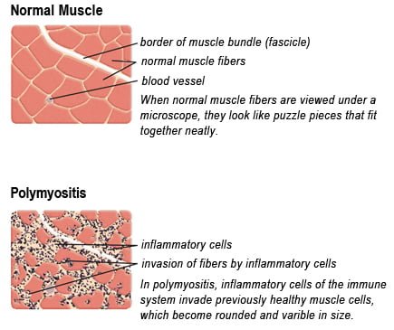
Drawing of Biopsy Showing Muscle Fibers Invaded by Immune Cells. Courtesy MDA.org
What Is Polymyositis?
P olymyositis is a body-wide connective tissue disease resulting from autoimmune attack of skeletal muscles that is characterized by inflammatory and degeneratory changes. The course is unpredictable being marked by spontaneous flare-ups and remissions.
Polymyositis can begin slowly or abruptly according to the factor that is triggering the onset such as infection, medications like phenytoin, and autoimmune disease.
Progressive muscle weakness starts in the proximal skeletal muscles (muscles closest to the trunk of the body).
Skeletal muscles, also called voluntary, are muscles that move the body as we want, such as walking and lifting objects, as opposed to those we cannot voluntarily control, such as the muscles of digestion.
Q: What are the degeneratory changes in skeletal muscles?
A: In polymyositis, degeneratory changes in skeletal muscles means that muscles are being destroyed (called necrosis), resulting in fibrosis, or scarring. When scar tissue takes the place of lost muscle tissue, it cannot act like muscle to contract and relax. Muscle destruction is what causes muscle pain and weakness.
After the clinical work-up of exams and blood studies to determine muscle damage, the diagnosis of polymyositis is confirmed by muscle biopsy. See image at above left. The black dots are inflammatory cells. Edema (fluid) between cells caused by inflammation pushes muscle fibers apart.
There is no cure for polymyositis, but the symptoms can be treated. Options include medication, physical therapy, exercise, heat therapy (including microwave and ultrasound), orthotics and assistive devices, and rest. The standard treatment for polymyositis is a corticosteroid drug, given either in pill form or intravenously. Immunosuppressant drugs, such as azathioprine and methotrexate, may reduce inflammation in people who do not respond well to prednisone.
Periodic treatment using intravenous immunoglobulin can also improve recovery. Other immunosuppressive agents used to treat the inflammation associated with polymyositis include cyclosporine A, cyclophosphamide, and tacrolimus. Physical therapy is usually recommended to prevent muscle atrophy and to regain muscle strength and range of motion.1
Diagnosis is based on elevated muscles enzymes, increased urinary creatine level, and electromyograph abnormalities.
Polymyositis can affect people at any age. It is most common in adults between ages 50 and 70, and in children ages 5 to 15. It affects women twice as often as men and is more common in African Americans than Caucasians.2 The major causes of death from polymyositis are cancer and lung disease, including pneumonia. The 5-year mortality rate can be as high as 1 in 5 patients.2
What Is Polymyositis In Celiac Disease and/or Gluten Sensitivity?
Sources:
National Institute of Neurological Disorders and Stroke [↩]