Skip to content

PMS (Premenstrual Syndrome) 

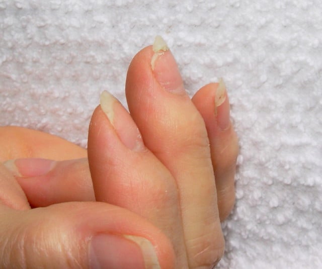
DSCN4758aWhat Are Brittle Nails?

B rittle nails are abnormalities of the nail plate that are characterized by poor nail structure affecting all fingernails and toenails seen as thin, dry nails that easily chip, split, and are difficult to maintain a clean edge. Usually longitudinal ridging occurs from the nail base to the tips.

Q: What is the nail plate?

A: The nail plate is the hard keratin cover protecting the finger tip and toe tip. The nail plate (non-living tissue) is produced by the nail matrix (living tissue) at the base of the nail plate under the lunula (moon), which is the site of brittle nail development.

Nail Anatomy. A. Nail plate; B. lunula; C. root; D. sinus; E. matrix; F. nail bed; G. hyponychium; H. free margin. Courtesy Wikipedia.org

Poor nail structure affecting all nails may be a feature of nutritional deficiency in poor diet or malabsorption such as occurs in celiac disease.

Some other causes are: idiopathic (unknown cause), the result of aging, the effects of certain drugs, or an association with systemic autoimmune disorders such as vitiligo, alopecia areata (with pitting), psoriasis (with pitting), and lichen planus. 

External (non-nutritional or disease) causes of dry, brittle nails, such as detergents and cleaners, would only affect fingernails but not toenails.

Note: It has been shown that working with your hands in water or soaking them through activities like swimming does not cause dry, brittle nails but will worsen them.

What Are Brittle Nails In Celiac Disease and/or Gluten Sensitivity?

Hypogonadism In Adult Males That Is Unexplained  

Nail Anatomy. A. Nail plate; B. lunula; C. root; D. sinus; E. matrix; F. nail bed; G. hyponychium; H. free margin. Courtesy Wikipedia.org
Nail Anatomy. A. Nail plate; B. lunula; C. root; D. sinus; E. matrix; F. nail bed; G. hyponychium; H. free margin. Courtesy Wikimedia.org

What Are Rounded Nails With Curved Ends?

R ounded, curved, dark and dry nails are a well known feature of vitamin B12 deficiency.

Q: How does vitamin B12 deficiency cause the nails to grow this way?

A: Vitamin B12 deficiency affects the nail plate. The nail plate is the hard keratin cover of the finger tip and toe tip, simply called “the nail.” The nail plate is produced by the living nail matrix at its base.

What Are Rounded Nails With Curved Ends In Celiac Disease and/or Gluten Sensitivity?

Impotence

This White Spot Is A Feature of Zinc Deficiency.

What Are White Spots And White Bands In Nails?

W hite spots in the nails is a feature of zinc deficiency and white bands signify protein deficiency characterized by abnormal appearance anywhere in one or more nails.

White spots and bands can appear separately or together in nails. There may be just one mark or many marks depending on the level of deficiency.

Q: What part of the nail is affected by zinc and/or protein deficiency?

A: Zinc and protein deficiencies affect the nail plate.

The nail plate is the hard keratin cover of the finger tip and toe tip, simply called “the nail.” The nail plate is produced by the living nail tissue at its base. Here is an illustration of the parts that make up a fingernail:

Nail Anatomy. Nail Anatomy. A. Nail plate; B. lunula; C. root; D. sinus; E. matrix; F. nail bed; G. hyponychium; H. free margin. Courtesy Wikipedia.org.

A. Nail plate; B. lunula; C. root; D. sinus; E. matrix; F. nail bed; G. hyponychium; H. free margin. Courtesy Wikipedia.

What Are White Spots And White Bands In Nails In Celiac Disease and/or Gluten Sensitivity?

Ocular Myopathy

What Is Primary Hyperparathyroidism?

Tissue changes in hyperparathyroidism. Courtesy Quizlet.com
Tissue Changes in Hyperparathyroidism Causing Brown Tumor. Courtesy Quizlet.com

P rimary hyperparathyroidism is a parathyroid disorder characterized by excessive secretion of parathyroid hormone by one or more parathyroid glands for more than 6 months.

In primary hyperparathyroidism, blood calcium levels are high while phosphorus levels are decreased due to the action of parathyroid hormone.

Parathyroid hormone is produced by the four pea sized parathyroid glands that are located on the thyroid gland in the front of the neck. Partly because the thyroid and parathyroid glands share the same anatomic place in the body and partly because they have similar names, they are often confused although they have completely different actions.

Parathyroid hormone normally keeps calcium and the opposing mineral phosphorus levels in balance by drawing calcium as needed from bones to increase it in blood and releasing excess phosphorus through the kidneys to decrease blood levels.

Primary hyperparathyroidism is commonly caused by an adenoma (tumor) in a parathyroid gland (80%) or 15% due to hyperplasia of gland tissue (overgrowth). It is seldom associated with autoimmune disorders. However, cancer is a  possibility.

Q: What is a parathyroid adenoma?

A: A parathyroid adenoma is usually a solitary, well circumscribed, soft, tan reddish-brown nodule with a capsule. Gland tissues outside of the adenoma are normal or slightly shrunken (not needed anymore).1

Untreated, primary hyperparathyroidism results in cyst formations in bone marrow (osteitis fibrosa cystica) and brown tumors in bone tissue. Cysts contain large amounts of fibrous tissue with areas of hemorrhage. Brown tumors contain aggregates of osteoclasts (bone cells), hemorrhage and giant cells resembling neoplasms.2

Here is the symptomatolgy: “Painful Bones, Renal Stones, Abdominal Groans, and Mental Moans.”

What Is Primary Hyperparathyroidism In Celiac Disease and/or Gluten Sensitivity?

Sources:
  1. http://quizlet.com/32428692/pathology-of-parathyroid-and-bone-flash-cards []
  2. http://quizlet.com/32428692/pathology-of-parathyroid-and-bone-flash-cards/ []

Infertility In Males

Cutaneous Vasculitis. Courtesy quizlet.com

What Is Cutaneous Vasculitis?

Cutaneous vasculitis is a vascular disease of small blood vessels serving the skin that is characterized by segmental (spotty) inflammation of affected vessel walls resulting in their necrosis (destruction). Scarring is seen in the affected skin that lies over the destroyed blood vessels.

Skin appearance varies from small flat, purplish spots to raised purplish areas over damaged vessels.

In most cases, cutaneous vasculitis results from deposits of toxic immune complexes in the affected vessel walls that is caused by autoimmune activity targeting small blood vessels.1

Cutaneous vasculitis can also result from infection, drug reactions, or malignancies.

Who is Affected in the general Population? Cutaneous vasculitis affects all ages.

The overall annual incidence of cutaneous vasculitis was 38.6 persons per million in 1998.2

What Is Cutaneous Vasculitis In Celiac Disease and/or Gluten Sensitivity?

Sources:
  1. Abenavoli L, Proietti I, Leggio L, Ferrulli A, Vonghia L, Capizzi R, Rotoli M, Amerio PL, Gasbarrini G, Addolorato G. Cutaneous manifestations in celiac disease. World J Gastroenterol. 2006 Feb 14;12(6):843-52. []
  2. Watts RA, Jolliffe VA, Grattan CE, Elliott J, Lockwood M, Scott DG. Cutaneous vasculitis in a defined population–clinical and epidemiological associations. J Rheumatol. 1998 May;25(5):920-4. []

Dysgeusia (Impaired Taste)

DyspareuniaWhat Is Autoimmune Thyroiditis (Hypothyroidism)?

A utoimmune thyroiditis, also called Hashimoto’s thyroiditis or Hashimoto’s Disease, is an autoimmune destruction of thyroid tissue characterized by insufficient thyroid hormone circulating in the body that causes formation of a goiter (enlarged thyroid gland) and hypothyroidism.

Hypothroidism refers to the condition of markedly reduced secretion of thyroid hormone. There are other causes of hypothyroidism besides Hashimoto’s thyroiditis.

Hashimoto’s thyroiditis is often associated with other autoimmune diseases such as celiac disease.

In Hashimoto’s thyroiditis, a profusion of antibodies are produced, which build up in the blood. Left untreated so that much of the thyroid gland is destroyed, this condition may progress to the very serious and life-threatening condition called myxedema.

Note: In myxedema, protein, electrolytes, and water abnormally accumulate in between cells which produce firm, inelastic puffy skin that is cool, dry, rough, scaly, and may appear yellow; in some people, areas such as the ankles become crusty with a look of tree bark. Many systemic changes develop shown by significant slowing of mental and physical functions. Please see below.

Q: What thyroid tissue is targeted for destruction?

A: In Hashimoto’s thyroiditis, high levels of autoantibodies target thyroglobulin and thyroid peroxidase, leading to inflammation and destruction of the thyroid gland. The resulting fibrosis or scarring of the gland results in lack of thyroid hormone production.

The thyroid gland consists of a large number of closed vesicles that contain a homogenous substance called colloid, which contains the thyroglobulin. Thyroglobulin is an iodine-containing protein secreted by the thyroid gland and stored within its colloid, from which the thyroid hormones thyroxine (T4) and triiodothyroinine (T3) are derived.1

T3 is the active hormone and is made from T4. Thyroid hormones affect metabolism, brain development, breathing, heart and nervous system functions, body temperature, muscle strength, skin dryness, menstrual cycles, weight, and cholesterol levels.

Thyroid hormone production is regulated by thyroid-stimulating hormone (TSH), which is made by the pituitary gland in the brain. Normally, when thyroid hormone levels in the blood are low, the pituitary releases more TSH. When thyroid hormone levels are high, the pituitary decreases TSH production.

Hashimoto’s disease, with or without the development of hypothyroidism, is treated with synthetic thyroxine, which is man-made T4. Health care providers prefer to use synthetic T4, such as Synthroid® (Levothyroxine), rather than synthetic T3, because T4 stays in the body longer, ensuring a steady supply of thyroid hormone throughout the day. The thyroid preparations made with animal thyroid are not considered as consistent as synthetic thyroid.2

What Is Autoimmune Thyroiditis In Celiac Disease and/or Gluten Sensitivity?

Sources:
  1. Taber’s Cyclopedic Medical Dictionary. 19th ed. F.A. Davis Company. Philadelphia, PA. []
  2. National Endocrine and Metabolic Diseases Information Service. []

Diabetic Instability 

Vertebrae. Courtesy FreePik.com

What Is Elevated Bone Alkaline Phosphatase?

E levated bone alkaline phosphatase (BALP) is a laboratory result that indicates an abnormal blood level of this bone enzyme.

A bone alkaline phosphatase blood level is one of the most frequently used biochemical markers of bone formation.

Q: Why is the purpose of  bone alkaline phosphatase?

A: Bone alkaline phosphatase is produced by bone cells called osteoclasts in normal bone maintenance for the purpose of breaking down old or damaged bone so that other bone cells called osteoblasts can fill in the excavated areas with new bone. This process keeps bone stong and healthy.

Elevated bone alkaline phosphatase shows that more bone is being broken down than is being replaced. It can be caused by hyperparathyroidism, bone tumors from cancer, and malnutrition.

What Is Elevated Bone Alkaline Phosphatase (BALP) In Celiac Disease and/or Gluten Sensitivity?

Lymphoma, B-Cell Non-Hodgkin’s

e3343bf0a689b5f974a852fa2e84a718What Is Laryngospasm?

L aryngospasm is an acute disorder of the larynx that is characterized by life-threatening spasms of the muscles of the larynx located in the throat.

Laryngospasm is a rare feature of hypocalcemia (low blood calcium level) characterized by severe alteration in nerve conduction and muscle contraction.

What Is Laryngospasm In Celiac Disease and/or Gluten Sensitivity?

Migraine (Headache)

Baby with Allergic Reaction to Peanuts. GFW
Baby with Allergic Reaction to Peanuts. GFW photo.

What Is Food Allergy?

F ood allergy is an abnormal immune response to food proteins that may damage the small intestinal  lining and produce malabsorption of food. The reaction is usually delayed which makes it difficult to identify the offending food that is causing symptoms.

Q: How does food allergy develop?

A: The gastrointestinal tract serves not only to digest and absorb foodstuffs but also to protect the body from unwanted substances. When allergic food substances are eaten, the immune response that is triggered in the gut produces inflammation with symptoms such as pain, vomiting and loose bowels.

Inflammation causes swelling of the gut lining that can interfere with the passage of nutrients through it to the body which results in malabsorption. Malabsorption deprives the body of nutrients needed for normal function.

Symptoms other than digestive may involve skin rashes, hives, and respiratory difficulties that can be distressing and life-threatening.

What Is Food Allergy In Celiac Disease and/or Gluten Sensitivity?

Celiac Disease, Refractory

Anatomy of Upper Airway and Throat. Courtesy Wikipedia.org

What Is Post-Cricoid Carcinoma?

P ost-cricoid carcinoma is a rare malignancy arising in the hypopharynx.

Q: What is the hypopharynx?

A: The hypopharynx is the lower portion of the pharynx that opens into the larynx. It is comprised of the postcricoid region, pyriform sinus, and posterior hypopharyngeal wall.

Tumors rarely appear in the posterior pharyngeal wall or postcricoid region without also involving the pyriform sinus. The lack of anatomic barriers between these sites and a propensity for these tumors to develop in the pyriform sinus and spread outward account for this phenomenon.

Overall prognosis for these tumors is poor. Because of the rich organic lymphatic and vascular networks, aggressive growth and early cervical metastases (compared with cancers at other head and neck sites) characterize these cancers.

A study investigating the clinical course of 21 patients with post-cricoid carcinoma found the 5-year cause-specific survival rate was 52%. All the patients who eventually died did so within 3 years of the treatment. Seven patients had distant metastases, representing a higher frequency as compared with that of patients with recurrence of the primary focus and cervical lymph node metastasis. All of these patients who had been treated by surgery died of the cancer.1

Unfortunately, hypopharyngeal cancers are discovered at a later stage than other head and neck cancers. The delay in diagnosis from symptom onset averages 10 months.2

What Is Post-Cricoid Carcinoma In Celiac Disease and/or Gluten Sensitivity?

Sources:
  1. Asakage T, Nakao K, Ebihara Y, Fujishiro Y, Watanabe K. A clinical study of post-cricoid carcinoma. Acta Otolaryngol Suppl. 2007 Dec;(559):118-22. doi: 10.1080/03655230701599354. []
  2. http://emedicine.medscape.com/article/848359-overview []