Skip to content

Candida Albicans Infection

Close-up shows inflammation and yellowish white patches of roof and back of the mouth caused by candidiasis. Uvula is greatly swollen, hanging near the level of the tongue. Courtesy: Wikipedia.
Close-up shows inflammation and infected patches on roof and back of the mouth caused by candidiasis. Uvula is swollen, hanging near the tongue. Courtesy: Wikimedia.

What Is Candida Albicans Infection?

Candida albicans infection, called candidosis or candidiasis, is an opportunistic invasion of mucous membrane or skin by candida albicans, an endogenous yeast found in 40 to 80% of normal human beings. A former name for this small, budding fungus is monilia albicans.

Opportunistic means that yeast living on mucosal and skin surfaces does not invade (infect) unless these tissues become unhealthy and therefore cannot protect themselves.

Q: How does candida albicans cause infection?

A: Candida albicans lives on the mucosal surfaces and skin in most people without causing infection (colonizes) because of our normal defenses against invasion. In fact, candida albicans is a very effective colonizer of humans. For example, Russell and Lay found that 47% of 1-month-old infants were orally colonized with candida albicans, and 49% were colonized with other fungi.

During growth within the intestinal tract, the organism senses pH (acidity), oxygen, carbon sources, and the presence of surfaces allowing it to optimize gene expression for a particular environment. With these mechanisms for sensing, candida albicans is able to efficiently colonize humans in infancy.1

Candida Infection Of The Esophagus on X-ray. Courtesy Radiology Assistant.nl
Candida Infection Of The Esophagus (White Area) On X-ray. Courtesy Radiology Assistant.nl

Lowered host defenses allow yeast already present on mucosal and skin surfaces to take advantage and can grow rapidly, becoming pathogenic (disease producing) so that infection results.

Infection is characterized by superficial, irregular white patches on mucosal surfaces and possible invasion of the bloodstream by a filamentous form (thread-like structures) that can rapidly develop.

Candida albicans is unique among oral pathogens in its ability to invade cornified layers of stratified squamous epithelium of the tongue, mouth surfaces, hard and soft palate, esophagus, and gut. Stratified squamous epithelium is the tough surface cells that ordinarily protect underlying tissues from damage or invasion by microbes.

Candida albicans is also capable of invading the lungs and causing pneumonia and septicemia, which is the spread of infection into the bloodstream.

Here is a time honored simple do-it-youself test for infection of the mouth or throat: First thing in the morning before brushing your teeth or eating, fill a small see through glass with water then gently spit onto the surface. If after an hour the spit remains on the water surface, it is unlikely you have candida in the mouth. If it grows legs downward, it indicates that yeast is growing. If the spit sinks to the bottom, you have this problem. Yeast in the mouth can quickly travel down the esophagus and into the gut.

Medical diagnosis. Difinitive diagnosis for the oral cavity is made by your clinician by swabbing the areas of your mouth and/or throat and viewing under a microscope for evidence of candida.

Infections of the esophagus and gut require inspection by gastroscopy or endoscopy procedure and the taking of samples to be examined under microscope. This examination also give the opportunity to rule out other problems. Barium swallow can show the extent of infection and any disfiguration of the esophagus that results.

What Is Candida Albicans Infection In Celiac Disease and/or Gluten Sensitivity?

Sources:
  1. Rosenbach A, Dignard D, Pierce JV, Whiteway M, Kumamoto CA. Adaptations of Candida albicans for growth in the mammalian intestinal tract. Eukaryot Cell. 2010 Jul;9(7):1075-86. doi: 10.1128/EC.00034-10. Epub 2010 Apr 30. []

Tuberculosis – Increased Susceptibility 

What Is Irritable Bowel Syndrome? I rritable bowel syndrome (IBS) is a motility disorder without anatomic cause involving the entire gastrointestinal tract that is characterized by these four features: 1) Abdominal pain usually relieved by… 

Iron Deficiency

ferritinWhat Is Iron?

Iron is an essential mineral that is required for normal body function.

Almost two-thirds of iron in the body is found in hemoglobin, the protein in red blood cells that carries oxygen to tissues. Smaller amounts of iron are found in myoglobin, a protein that helps supply oxygen to muscle, and in enzymes that assist biochemical reactions.

Iron is also found in proteins that store iron for future needs and that transport iron in blood.

Q: How are iron stores regulated?

A:Iron stores are regulated by intestinal iron absorption.1That is, whenever iron is needed, more iron is absorbed than when iron is sufficient. This mechanism prevents excess iron in the body which is harmful.

In the brain, iron is necessary to ensure oxygenation and to produce energy in the cerebral parenchyma (via cytochrome oxid. ase), and for the synthesis of neurotransmitters and myelin.

Iron concentrations in the umbilical artery are critical during the development of the fetus, and in relation with the IQ in the child.2

What Is Iron Deficiency In Celiac Disease and/or Gluten Sensitivity?

Sources:
  1. http://ods.od.nih.gov/factsheets/Iron-HealthProfessional accessed 12/11/12 []
  2. Bourre JM. Effects of nutrients (in food) on the structure and function of the nervous system: update on dietary requirements for brain. Part 1: micronutrients. J Nutr Health Aging. 2006 Sep-Oct; 10(5):377-85. []

Sarcoidosis

What Is Vitamin B12 Deficiency Anemia? V itamin B12 deficiency anemia is a megaloblastic anemia that is characterized by defective DNA synthesis of red blood cells due to a lack of vitamin B12. Vitamin B12 is… 

Pulmonary Hemosiderosis, Idiopathic  (Lane-Hamilton Syndrome)

intest (2)What Is Obnoxious Gas?

O bnoxious gas, or flatus, is gas that is not only offensive when passed but also lingers in the air longer than ordinary gas does.

Gas is a natural digestive product within the colon that is composed mainly of hydrogen and carbon dioxide gases. These gases are given off in the necessary bacterial breakdown of undigested fermentable food entering the colon from the small intestine.

Q: What makes gas obnoxious?

A: The production of obnoxious gas depends on the type and quantity of undigested food residue that is passed into the colon from the small intestine, dysfunctional motility instead of normal peristalsis, and dysbiosis. Dysbiosis is the condition of  having unhealthy or insufficient populations of microbes responsible for digesting (fermenting) foodstuffs in the lower gut or colon. 

Gases produced by intestinal microbes may modulate intestinal motor function (muscle movement) in individuals with functional bowel disease. Methane, produced by enteric bacteria in the human gut, is associated with slowed intestinal transit and constipation.1

What Is Obnoxious Gas In Celiac Disease and/or Gluten Sensitivity?

Sources:
  1. Jahng J, Jung IS, Choi EJ, Conklin JL, Park H. The effects of methane and hydrogen gases produced by enteric bacteria on ileal motility and colonic transit time. Neurogastroenterol Motil. 2012 Feb;24(2):185-90, e92. doi: 10.1111/j.1365-2982.2011.01819.x. Epub 2011 Nov 20.

    Methane is produced in the colon by intestinal methanogens (microbes) that metabolize hydrogen, one of the end products of normal anaerobic (meaning without oxygen) bacterial fermentation.  Fermentation of the undigested starchy part of carbohydrates produces hydrogen in the intestine, which is the food for methane production by intestinal methanogens.

    Hydrogen and methane are excreted in the flatus and in breath giving the opportunity to indirectly measure their production using breath testing. ((Triantafyllou K, Chang C, Pimentel M. Methanogens, Methane and Gastrointestinal Motility. J Neurogastroenterol Motil. 2014 Jan;20(1):31-40. Epub 2013 Dec 30. []

Bronchiectasis

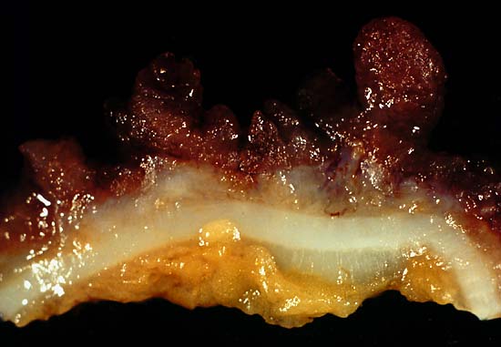
This photo was from a total colectomy done for clinically severe, intractable chronic ulcerative colitis. It shows a closer view of a longitudinal section through the colon wall. This demonstrates not only the angry red mucosa but also the tendency for the inflamed tissue to throw itself up into inflammatory pseudopolyps. Source: Ed Uthman, MD. Public domain.
This photo is from a total colectomy done for severe, intractable chronic ulcerative colitis. It shows a close view of a lengthwise section through the colon wall. This demonstrates not only the angry red mucosa, but also, the tendency for the inflamed tissue to throw itself up into inflammatory pseudopolyps.
Source: Ed Uthman, MD. Public domain.

What Is Ulcerative Colitis?

U lcerative colitis is an inflammatory disorder of the colon characterized by continuous inflammation of the mucosa and submucosa usually with small ulcers, extending from the rectum and typically involving the distal colon, rectum, and anus and producing bloody diarrhea.

While the severity of ulcerative colitis varies among patients, iron deficiency anemia often develops due to blood loss especially when there are many bloody bowel movements in a day.

The onset of ulcerative colitis is most commonly in young adulhood.

Q: Is this disease painful?

A: Yes with the passage of stool.

Psychological stress and subsequent dysbiosis exacerbate ulcerative colitis.

Psychological stress activates multiple physiological processes aimed at maintaining balance within the body. These physiological processes also have the capacity to influence the composition of microbial communities in the digestive tract, and research now indicates that exposure to stressful stimuli leads to gut microbiota dysbiosis.1

While the relative abundance of many different bacterial types can be altered during stressor exposure, findings in nonhuman primates and laboratory rodents, as well as humans, indicate that bacteria in the genus Lactobacillus are consistently reduced in the gut during stress.2

Presently, ulcerative colitis cannot be cured. This condition has a course of remissions, when symptoms subside, and flares, when symtpoms get worse. Treatment is aimed to reduce flares and promote remission. In all cases, correction of dysbiosis improves the condition.

Ulcerative colitis is associated with increased incidence of cancer of the colon.3

What Is Ulcerative Colitis In Celiac Disease and/or Gluten Sensitivity?

Sources:
  1. Galley JD, Bailey MT. Impact of stressor exposure on the interplay between commensal microbiota and host inflammation. Gut Microbes. 2014 May 1;5(3):390-396. Epub 2014 Apr 1. []
  2. Galley JD, Bailey MT. Impact of stressor exposure on the interplay between commensal microbiota and host inflammation. Gut Microbes. 2014 May 1;5(3):390-396. Epub 2014 Apr 1. []
  3. Taber’s Cyclopedic Medical Dictionary. F. A. Davis. Philadelphia, PA []

Asthma

Depiction of pancreatic amylase molecule.

What Is Macroamylasemia?

M acroamylasemia is an acquired enzyme disorder that causes elevated levels of the enzyme amylase (hyperamylasaemia) in the bloodstream. It is characterized by altered amylase molecules that have become abnormally bound with plasma proteins in the bloodstream, commonly IgG (immunoglobulin G) and/or IgA (immunoglobulin A).

The resulting molecule is too large to be properly filtered by the kidneys and excreted in the urine as would be normal unbound amylase, causing sustained elevation of amylase levels in the plasma.

With normal kidney function, a hyperamylasemia without an increase in urine amylase suggests the diagnosis of macroamylasemia, and is confirmed by identifying the macromolecular components.1

Amylase is a an enzyme produced by the pancreas and the parotid glands to digest starch in the diet. Comparatively small amounts are also produced by other organs.

Q: How do the pancreas and parotid glands deliver their amylase for digestion of starch?

A: The parotid glands deliver their amylase into the mouth. The action of amylase in the mouth starts the breakdown of starch as it is chewed and moistened with saliva.

After the starchy food is liquified in the stomach, the mass empties into the duodenum. Here pancreatic amylase is delivered through the common bile duct into the duodenum where it turns starch into maltose sugar. The next step to final digestion of starch is carried out by enzymes in the small intestinal lining that split maltose into its component sugar which is glucose.

Glucose is a simple sugar that can be absorbed into the bloodstream for use in the body. Humans cannot live without adequate glucose.

Distinquishing macroamylasemia from hyperamylasemia due to pancreatic disease is necessary to prevent needless treatment and investigation for pancreatitis.2

What Is Macroamylasemia In Celiac Disease and/or Gluten Sensitivity?

Sources:
  1. Torrent Vernetta A, Segarra Cantón O, Soler Palacín P, Segura Cardona RM, Infante Pina D. Macroamylasaemia in paediatrics. An Pediatr (Barc). 2008 Nov;69(5):439-41. []
  2. Isham CA, Ridgeway NA, Hedrick R, Cate JC 4th. Screening for macroamylase in a community hospital. Clin Chem. 1984 May;30(5):741-2. []

Tuberculosis – Non-Response to Treatment 

Underarm showing skin darkening, which is a feature of Addison's Disease
Underarm showing skin darkening, which is a feature of Addison’s Disease

What Is Primary Addison’s Disease?

A ddison’s disease is an autoimmune destruction of the adrenal glands by autoantibodies that target the adrenal cortex, or outer part of these glands, and is characterized by a slow progressive failure of the adrenal glands to adequately produce its steroid hormones.

Symptoms of adrenal fatigue or failure may not develop until the majority of adrenal tissue is destroyed. When untreated, progression leads to coma, called Addisonian crisis, which is a medical emergency.

There are two adrenal glands each located on top of a kidney and enclosed in a connective tissue capsule. Each is a small, triangular shape that is made of two parts: the outer region and the inner region.

The inner region, called the adrenal medulla, produces epinephrine and norepinephrine chemicals that are needed to deal with stress.

The outer region, called the adrenal cortex, produces adrenocortical (steroid) hormones and releases them into the bloodstream in response to pituitary stimulating hormone from the brain.

Q: What is the function of steroid hormones produced by the adrenal glands?

A: Functions of the three steroid hormones produced by the adrenal glands are:

  1. Glucocorticoids restrain inflammation and metabolism of carbohydrates, fats and proteins to maintain a normal glucose blood level. The major glucocorticoid is hydrocortisone.
  2. Mineralocorticoids regulate the retention and excretion of fluids and electrolytes by the kidneys. The most important mineralocorticoid is aldosterone.
  3. Androgen (testosterone) is a male sex hormone.

Secondary adrenal insufficiency may develop from other causes that are not immune related such as chronic infections, tumor, and medications.

What Is Addison’s Disease In Celiac Disease and/or Gluten Sensitivity?

Pulmonary Permeability, Increased 

Erosions Of The Esophagus Can Be A Cause Of Gastrointestinal Bleeding.
Erosions Resulting From Esophagitis Can Be A Cause Of Gastrointestinal Bleeding.

What Is Occult Gastrointestinal Bleeding?

O ccult gastrointestinal bleeding is characterized by unseen or minute quantities of blood in stool. The origin of bleeding is from mucosa that lines the inside of the digestive tract at a site that must be found by medical testing and procedures to look at the lining.

Q: What tests and procedures are performed to locate the bleeding?

A: The simplest test to discover blood that cannot be seen is the stool test. This consists of a card with 3 spaces for 3 separate  stool collection over 3 separate days. A tiny smear of stool is placed on a test card space on each of three days. Then the card is examined for a reaction that indicates the existence of blood in the stool.

If the stool test is positive, the origin of bleeding must be located. This search requires certain procedures that look at the mucosal lining directly by a gastroenterologist, usually under light sedation.

  • Gastroscopy procedure can visualize the upper gastrointestinal tract, which includes the esophagus and stomach.
  • Endoscopy procedure can also visualize the upper small intestine. However, endoscopy procedure is limited in that it cannot visualize the middle small intestine (jejunum).
  • Colonoscopy can visualize the end of the small intestine and colon all the way to the rectum.
  • Capsule endoscopy procedure.  If  gastroendoscopy and colonoscopy fail to discover the source of bleeding, the physician may administer a capsule endoscopy which visualizes the entire gastrointestinal tract. In this procedure, the patient swallows a capsule size camera (encased) which takes constant pictures over the course of a day until expelled through the rectum.  Of course, the patient must be able to swallow the large size capsule.

What Is Occult Gastrointestinal Bleeding In Celiac Disease and/or Gluten Sensitivity?

Abscess Formation Causing Lung Cavities

Bristol Stool Chart Showing Normal and Abnormal Stool.

What Is Chronic Constipation Alternating With Diarrhea?

C hronic constipation alternating with diarrhea is an intestinal motility disorder, or irregularity, characterized by alteration in stool formation, consistency, and evacuation which results in a bowel movement that consists of some hard or balled stool along with some loose stool that can cause leakage.

Q: How do irregular movement patterns develop in the colon?

A: The colon produces irregular movements as a result of problems that originate in  the colon (large intestine) itself and/or the small intestine which then affects function of the colon.

Here are listed the many types of problems or diseases that cause these abnormal bowel movements:

  • Disorders that adversely affect the colon, an organ which must propel stool, remove excess water, absorb electrolytes, ferment undigested food material that passes into it, and produce nutrients from the fermentation process:
  • Poor diet that does not contain adequate nutrition, fiber, probiotics, prebiotics, and water to form normal stool.
  • Diet that contains irritating, toxic or allergenic food that cause spasms.
  • Diseases that inflame the mucosa lining such as collagenous colitis, altering the proper absorption of water and electrolytes.
  • Diseases that damage and swell the colon walls, such as Crohn’s disease, ulcerative colitis, and diverticulitis.
  • Diseases that obstruct the lumen or passageway so that stool passes with difficulty.
  • Diseases that hamper normal peristalsis (muscle action), such as irritable bowel syndrome (IBS), diabetes and thyroid disease.
  • Disorders that adversely affect the small intestine, an organ which must digest and absorb nutrients needed by the body while passing unabsorbed food material to the colon:  
  • Diet that conatins too much fat, sugar or artifical sweeteners, causing diarrhea.
  • Disorders that result in malabsorption, such as gluten enteropathy, milk enteropathy, steatorrhea (fat malabsorption), lactose intolerance, sucrose intolerance, maltose intolerance, and bacterial overgrowth, passing abnormal amounts of undigested food material to the colon where it is fermented producing excessive gas, diarrhea and spasm.
  • Disorders that impair peristalsis, such as active celiac disease, diabetes, scleroderma, and thyroid disease.
  • Tumors like cancer and lymphoma impair regular passage of material to colon.
  • Drugs that impair peristalsis, such as iron supplements, aluminum containing antacids, narcotics, some anti-depressants, some anti-seizure, and some diuretics.

What Is Chronic Constipation Alternating With Diarrhea In Celiac Disease and/or Gluten Sensitivity?