Skip to content

Obstetrical Complications

eczemaWhat Is Eczema?

E czema, or atopic dermatitis, is a chronic recurring inflammatory skin disorder characterized by itching, eczematous plaques and a defective epidermal barrier (surface layer of skin cells) that fails to hold moisture, with affected areas becoming dry then inflamed.

Q: Why does eczema develop?

A: Patients with atopic dermatitis have genetically determined risk factors that affect the barrier function of the skin and immune responses that interact with environmental factors.1

Atopic eczema is associated both with an abnormal skin matrix and impaired systemic immune response. Therefore, isolated topical treatment may have suboptimal effect.2 

Atopic eczema is a common presentation of  immune response towards food proteins in infancy.3

Global evidences reflect a marked increase in prevalence, which has tripled since 1960. In the United States, the current prevalence rates range from 10% to 20% in children and 1 to 3% in adults.4

What Is Eczema In Celiac Disease and/or Gluten Sensitivity?

Sources:
  1. Mesquita Kde C, Igreja AC, Costa IM. Atopic dermatitis and vitamin D: facts and controversies. An Bras Dermatol. 2013 Nov-Dec;88(6):945-53. doi: 10.1590/abd1806-4841.20132660. []
  2. Caffarelli C, Santamaria F, Vottero A, Bernasconi S. Progress in Pediatrics in 2012: choices in allergy, endocrinology, gastroenterology, hematology, infectious diseases, neurology, nutrition and respiratory tract illnesses. Ital J Pediatr. 2013 May 8;39(1):26. []
  3. Ho MH, Wong WH, Chang C. Clinical Spectrum of Food Allergies: a Comprehensive Review. Clin Rev Allergy Immunol. 2012 Nov 16. []
  4. Mesquita Kde C, Igreja AC, Costa IM. Atopic dermatitis and vitamin D: facts and controversies. An Bras Dermatol. 2013 Nov-Dec;88(6):945-53. doi: 10.1590/abd1806-4841.20132660. []

Miscarriage (Spontaneous Abortion)

Pitting edema. Right photo shows that indent remains from pressing. Courtesy wikimedia.

What Is Edema?

E dema is an abnormal swollen condition of the skin characterized by excess extracellular fluid volume, meaning there is an increase of the fluid that normally surrounds cells. Edema may be hardly noticeable or it can become extensive.

Edema can have various appearances and can develop from various causes.

Q: What are the appearances and causes of edema?

A: Here are the appearances of edema:

  • Pitting edema. It is called pitting edema if when the skin is pressed with a finger, the indent remains. This edema results from fluid leaking out of the bloodstream into the surrounding tissues. Pitting edema can be a feature of many disorders including heart disease, kidney disease, vascular disease, cancer, and malnutrition.
  • Myxedema is a firm swelling or thickening of subcutaneous skin that does not pit, as seen in thyroid disease.

Here are causes of edema:

  • Local edema as a response to trauma or infection.
  • Lower extremity edema from poor circulation and malnutrition.
  • Edema in the lungs from right sided heart disease.
  • Abdominal edema from liver disease, intestinal disease.
  • Body wide edema from heart disease, kidney disease, thyroid disease.

What Is Edema In Celiac Disease and/or Gluten Sensitivity?

Short Duration of Breast Feeding 

dermatomyositisWhat Is Dermatomyositis?

D ermatomyositis is a rare autoimmune systemic disease of the connective tissue that is characterized by inflammatory and debilitating degenerative changes in the muscles and in the skin. 

Dermatomyositis results in symmetric, proximal muscle weakness of limbs (upper arms and legs), and skin manifestations. 50-70% of patients have circulating myositis-specific auto-antibodies.

The course of dermatomyositis is unpredictable being marked by spontaneous flare-ups and remissions. It can begin slowly or abruptly according to the factor that is triggering the onset such as infection, medications like phenytoin, and autoimmune disease.

Q: What are the skin manifestations of dermatomyositis?

A: Classic skin manifestations of dermatomyositis include these features:

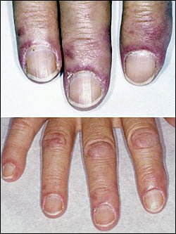
  • The heliotrope rash (lilac color) on upper eyelids.
  • Rash on face, neck, shoulders, upper chest, elbows, knees, knuckles, and back.
  • Gottron’s papules (scaly, red eruptions or  patches over the knuckles, elbows, and knees).
  • The V-sign (rash front of neck and chest).
  • The shawl sign (rash distribution on shoulders and back).1

Additional cutaneous manifestations are described below under symptoms.

Dermatomyositis is associated with an increased risk of  cancer, other autoimmune diseases, such as lupus and psoriasis, and it can be a complication of interferon-α therapy. About 1 person in 100,000 are affected according to various studies. While it affects all ages, women have twice the occurence of men.

There is no cure for dermatomyositis, but the symptoms can be treated.  Options include medication, physical therapy, exercise, heat therapy (including microwave and ultrasound), orthotics and assistive devices, and rest.  The standard treatment for dermatomyositis is a corticosteroid drug, given either in pill form or intravenously.  Immunosuppressant drugs, such as azathioprine and methotrexate, may reduce inflammation in people who do not respond well to prednisone.2

What Is Dermatomyositis In Celiac Disease and/or Gluten Sensitivity?

Sources:
  1. Marvi U, Chung L, Fiorentino DF. Clinical presentation and evaluation of dermatomyositis. Indian J Dermatol. 2012 Sep;57(5):375-81. doi: 10.4103/0019-5154.100486. []
  2. National Institute of Neurological Disorders and Stroke. []

Anemia, Severe Iron Deficiency In Pregnancy 

Dermatitis Herpetiformis. Blisters Opened Giving Relief From Pain and Itching.
Dermatitis Herpetiformis On Forearm. Skin Is Darkened Where Old Blisters Healed.

What Is Dermatitis Herpetiformis?

D ermatitis herpetiformis (DH) is an autoimmune extremely itchy, painful bullous skin rash (blistering eruptions) arising from the underlying dermis layer of skin as a consequence of gluten sensitivity.

Dermatitis herpetiformis is characterized by multiple intensely itchy, red blisters appearing on the elbows which can extend down the forearm to the wrist and the knees. Less usual areas involve the back, buttocks, scalp, and abdomen.

Q: Do the blisters leave a mark when healed?

A: Crops of skin eruptions begin with itching or a burning sensation in reddened papules. There are grouped vesicles and tense blisters. The blister contents may be serous or bloody, with symmetrical distribution (eg, both knees or both elbows). Fluid filled elements rupture leaving denuded areas of sore skin and crust. Subsequently, there is residual hypopigmentation (a white area) or hyperpigmentation (dark area).1

Rupture of blisters begins relief from intense burning and itching.

Dermatitis Herpetiformis Eruptions On Knees.
Dermatitis Herpetiformis Eruptions On Knees. Notice White Areas Showing Loss of Pigmentation From Healed Blisters.

What Is Dermatitis Herpetiformis In Celiac Disease and/or Gluten Sensitivity?

Primary care providers should be aware of this skin condition, as they are more likely than a gastroenterologist to be confronted with this type of presentation of celiac disease.2

Sources:
  1. Mendes FB, Hissa-Elian A, de Abreu MA, Gonçalves VS. Review: dermatitis herpetiformis. An Bras Dermatol. 2013 Jul-Aug;88(4):594-9. []
  2. Robinson BL, Davis SC, Vess J, Lebel, J. Primary care management of celiac disease. Autoimmune Disorders. Nurse Practitioner. February 2015: Vol 40 – Issue 2; 28–34. []

Celiac Disease After Childbirth (Puerperium)  

erythema elevatum diutinum of forearmWhat Is Erythema Elevatum Diutinum?

E rythema elevatum diutinum is a rare leucocytoclastic vasculitis thought to be caused by an immune response that is characterized by inflammation, a rash, and scarring of small blood vessels of the skin.

A characteristic poorly defined rash develops symmetically in the skin of extremities where the vessels are inflamed. Nodules and plaques are yellowish red and crust on healing, leaving darkened areas (hyperpigmentation).

Q: What is leucocytoclastic vasculitis?

A: Leucocytoclastic vasculitis is the most common form of vasculitis (inflammation of blood vessels) of the skin and usually results from deposition of immune complexes at the vessel wall. It presents in different forms and in association with different diseases. Diagnosis is made by skin biopsy with immunofluorescence.1

Erythema elevatum diutinum may be caused by inflammatory disease, certain infections such as HIV, immunological disease, malignancy, and drugs.

Corticosteroids are indicated when there are signs of incipient skin necrosis or when the disease is severe. Otherwise, treatment is with Colchicine or Dapsone medications.2

What Is Erythema Elevatum Diutinum In Celiac Disease and/or Gluten Sensitivity?

Sources:
  1. Sunderkötter C, Bonsmann G, Sindrilaru A, Luger T. Management of leukocytoclastic vasculitis. J Dermatolog Treat. 2005;16(4):193-206. []
  2. Sunderkötter C, Bonsmann G, Sindrilaru A, Luger T. Management of leukocytoclastic vasculitis. J Dermatolog Treat. 2005;16(4):193-206. []