Skip to content

Progressive Myoclonic Ataxia 

biliary system primary biliary cirrhosisWhat Is Primary Biliary Cirrhosis?

P rimary biliary cirrhosis (PBC) is a biliary tract disease characterized by chronic cholestasis (build-up of bile) and gradual destruction of bile ducts within the liver, called intrahepatic bile ducts, caused by chronic inflammation.

Primary biliary cirrhosis comes under the umbrella term autoimune liver disease in which the end result is immune-mediated hepatocellular (liver cell) or hepatobiliary (bile duct) injury.1

Q: What is the end result of destruction of bile ducts?

A: The end result of destruction of bile ducts is liver damage.

Injured liver tissue from chronic inflammation and the buildup of bile leads to cirrhosis, a condition in which the liver slowly deteriorates and malfunctions.

Scar tissue replaces healthy liver tissue, partially blocking the flow of blood through the liver. Scarring also impairs the liver’s normal ability to control infections, remove bacteria and toxins from the blood, process nutrients, hormones, and drugs, make proteins that regulate blood clotting, produce bile, and effectively replace its own cells when they become damaged.2

The liver is the largest organ within the body. It lies mostly in the upper part of the abdomen on the right side just under the diaphragm. About 70% of liver tissue is made up of cube shaped cells called hepatocytes that do the main work of the liver. Other cells (epithelial) form structure and are arranged in single layers around blood vessels, sinusoids, and bile ducts.

Bile ducts carry bile, a greenish brown liquid made by the liver to the gall bladder for storage until needed to aid in the digestion and absorption of fat and fat-soluble vitamins A, D, E, and K from the small intestine. Bile emulsifies fat eaten in the diet so that the pancreatic enzyme called lypase can break it down into its fatty acid and glycerol components which can then be absorbed into the body.

Bile also carries away waste products produced by normal metabolism and toxic substances that are removed by the liver for eventual elimination in stool. Bile is continually made by the liver from phospholipids, bile acids, cholesterol, and aging blood cells it removes from circulation. As such, bile must continually flow out of the liver to prevent build-up in the liver.

There is no cure for primary biliary cirrhosis.

What Is Primary Biliary Cirrhosis In Celiac Disease and/or Gluten Sensitivity?

Sources:
  1. Trivedi PJ, Adams DH. Mucosal immunity in liver autoimmunity: a comprehensive review. J Autoimmun. 2013 Oct;46:97-111. doi: 10.1016/j.jaut.2013.06.013. []
  2. http://digestive.niddk.nih.gov/ddiseases/pubs/primarybiliarycirrhosis/ []

Congenital Anomalies 

Hangnail on lowest finger. Courtesy Wikipedia.org

What Is Hangnail?

H angnail is a broken strip of epidermis (piece of skin) at root or lateral (side) edge of fingernail or toenail that causes sharp pain.

A hangnail develops because the skin around the nail is unhealthy due to inadequate nutrition. Injury from trauma including biting the skin and pushing back the cuticles or exposure to excessive detergents and water that remove protective oils promote the development of hangnail.

All ages and both sexes can be affected.

Q: Can a hangnail become infected?

A: Infection, called paronychia, may develop from invasion of sore skin by any of these pathogens: bacteria, fungus, or yeast (Candida). Infected skin is red, swollen, and painful. Topical ointment is required to treat the infection.1

What Is Hangnail In Celiac Disease and/or Gluten Sensitivity?

Sources:
  1. www.nlm.nih.gov/medlineplus/ency/article/001444.htm []

Irritability

Small Bowel Volvulus, Courtesy  African Journal of Pediatric Surgery.
Small Bowel Volvulus Caused the  Enlarged Dark Loops. Courtesy African Journal of Pediatric Surgery.

What Is Volvulus?

V olvulus is twisting of a loop(s) of bowel onto itself which effectively closes it thus preventing digested matter from passing and causing engorgement of the closed loop with gas and fluid.

Q: How does the bowel get untwisted?

A: This condition usually necessitates surgical correction although some do return to their proper position.

The danger is that there may develop necrosis of the twisted loop(s) in which case, if left untreated, death will ensue.

What Is Volvulus In Celiac Disease and/or Gluten Sensitivity?

PMS (Premenstrual Syndrome) 

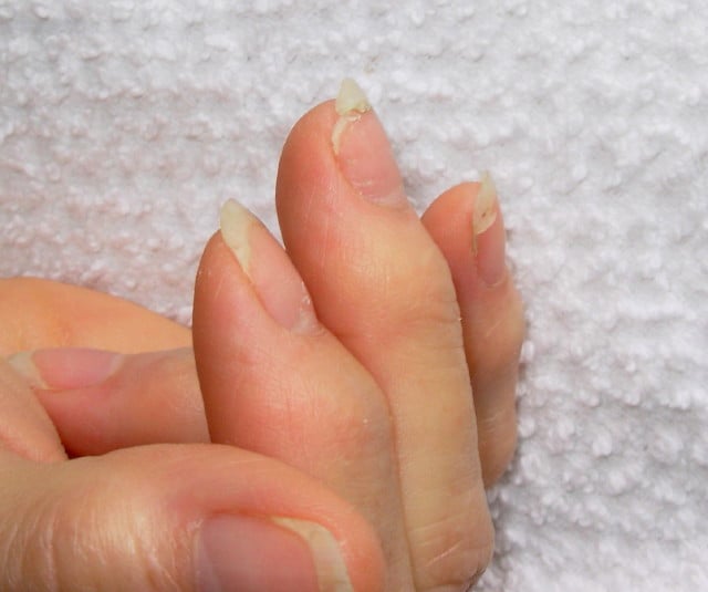
DSCN4758aWhat Are Brittle Nails?

B rittle nails are abnormalities of the nail plate that are characterized by poor nail structure affecting all fingernails and toenails seen as thin, dry nails that easily chip, split, and are difficult to maintain a clean edge. Usually longitudinal ridging occurs from the nail base to the tips.

Q: What is the nail plate?

A: The nail plate is the hard keratin cover protecting the finger tip and toe tip. The nail plate (non-living tissue) is produced by the nail matrix (living tissue) at the base of the nail plate under the lunula (moon), which is the site of brittle nail development.

Nail Anatomy. A. Nail plate; B. lunula; C. root; D. sinus; E. matrix; F. nail bed; G. hyponychium; H. free margin. Courtesy Wikipedia.org

Poor nail structure affecting all nails may be a feature of nutritional deficiency in poor diet or malabsorption such as occurs in celiac disease.

Some other causes are: idiopathic (unknown cause), the result of aging, the effects of certain drugs, or an association with systemic autoimmune disorders such as vitiligo, alopecia areata (with pitting), psoriasis (with pitting), and lichen planus. 

External (non-nutritional or disease) causes of dry, brittle nails, such as detergents and cleaners, would only affect fingernails but not toenails.

Note: It has been shown that working with your hands in water or soaking them through activities like swimming does not cause dry, brittle nails but will worsen them.

What Are Brittle Nails In Celiac Disease and/or Gluten Sensitivity?

Cortical Calcifying Angiomatosis

What Is Carbohydrate Malabsorption? C arbohydrate malabsorption is a digestive disorder characterized by the inability to properly digest and absorb carbohydrates within the small intestine to supply needed energy to the body. Q: What carbohydrates should be… 

Epilepsy (Convulsions)

chronic diarrhea gluten celiac disease symptomWhat Is Chronic Diarrhea?

C hronic diarrhea is an intestinal motility disorder characterized by 1) alteration in stool formation causing loose to fluid movements and quantity of movements with or without abdominal pain and 2) interference with normal carbohydrate salvage by the intestinal microbe population.

The severity of diarrhea is determined by the frequency and quantity of fluid lost.

Q: What is carbohydrate salvage by microbes in the colon?

A: Carbohydrate salvage in the colon is the necessary process whereby billions of microbes normally present in the colon work to breakdown undigestible carbohydrate foodstuffs such as fiber that continually arrive from the small intestine.

The microbial action releases energy to the body that would otherwise be lost with defecation, generates short-chain fatty acids, and stimulates sodium and fluid absorption. In this important process, butyrate and proprionate are produced which nourish the colonocytes (cells that line the colon), and acetate is produced for the liver.

While diarrhea may be a common symptom of small bowel mucosal disease, the consequent malabsorption can lead to substantial malnutrition and nutrient deficiencies. The small intestine, unlike the colon, has been relatively inaccessible, and systematic evaluation is often necessary to identify and treat small intestinal mucosal diseases that lead to diarrhea. All patients with severe diarrhea or diarrhea associated with features suggestive of malabsorption may have a disease of the small intestinal mucosa that requires careful evaluation and targeted management.1

What Is Chronic Diarrhea In Celiac Disease and/or Gluten Sensitivity?

Sources:
  1. Murray JA1, Rubio-Tapia A. Diarrhoea due to small bowel diseases. Best Pract Res Clin Gastroenterol. 2012 Oct;26(5):581-600. doi: 10.1016/j.bpg.2012.11.013. []

Migraine (Headache)

Baby with Allergic Reaction to Peanuts. GFW
Baby with Allergic Reaction to Peanuts. GFW photo.

What Is Food Allergy?

F ood allergy is an abnormal immune response to food proteins that may damage the small intestinal  lining and produce malabsorption of food. The reaction is usually delayed which makes it difficult to identify the offending food that is causing symptoms.

Q: How does food allergy develop?

A: The gastrointestinal tract serves not only to digest and absorb foodstuffs but also to protect the body from unwanted substances. When allergic food substances are eaten, the immune response that is triggered in the gut produces inflammation with symptoms such as pain, vomiting and loose bowels.

Inflammation causes swelling of the gut lining that can interfere with the passage of nutrients through it to the body which results in malabsorption. Malabsorption deprives the body of nutrients needed for normal function.

Symptoms other than digestive may involve skin rashes, hives, and respiratory difficulties that can be distressing and life-threatening.

What Is Food Allergy In Celiac Disease and/or Gluten Sensitivity?

Occipital Lobe Epilepsy With Cerebral Calcifications

Close-up Slice of a Small Intestinal Villus Showing How Enterocytes Appear Tightly Lining the Entire Outside Surface Of A Villus. Courtesy Cleo Libonati

What Is Increased Intestinal Permeability?

I ncreased intestinal permeability is characterized by dysfunctional intestinal permeability (leakiness) allowing for the penetration of harmful entities from the gut into the bloodstream such as undigested proteins and microbes. The popular name is “leaky gut.”

Q: Why does intestinal permeability increase?

A: Intestinal permeability is an essential function of the small intestinal mucosal lining by which wanted substances such as properly digested foodstuffs are allowed to permeate through the lining to enter the body via the bloodstream and lymphatics. At the same time unwanted substances are kept out.

The mucosal lining is one cell thick and makes up the surface between the digested foodstuffs inside the hollow of the intestine and the underlying tissues.

The mucosal lining is covered by millions of microscopic finger-like structures called villi that project toward the inside of the intestine giving the appearance of a shag rug.

Each one, called a villus, contains a capillary bringing blood to absorb nutrients, a vein to take away nutrients, and a lacteal to absorb and take away digested fat. Its wall is made up of a single layer of tightly connecting cells, called enterocytes.

This single layer of cells separates the contents of our small intestine from the lamina propria (underlying tissues of the small intestine) and the rest of our body. Breaching of this single layer of cells by leakiness can expose lymphocytes (immune cells) located in the lamina propria to a myriad of microorganisms and food antigens, leading to immune reactions.1

To protect the body from unwanted substances, a gatekeeping barrier system operates to regulate the passage of nutrients, or permeation, through the surface mucosal lining. This system acts to seal the inside body from the gut.

The integrity of intestinal permeability is determined by interactions among several barrier components including the unstirred water layer, mucosal surface hydrophobicity, the surface mucous coat, and cell factors (especially tight junctions).

Tight junctions hold cells tightly together side-by-side to prevent unwanted substances from passing through the lining. Tight junctions are complex structures comprising over 50 proteins, such as the claudin proteins which are considered to be the structural backbone of tight junctions.

Tight junctions include a series of special proteins forming fibrils (springy like proteins) that cross the plasma membrane and interact with proteins in the adjoining cells. Tight junctions are regulated by the protein zonulin.2

If zonulin deregulates from the action of substances such as gliadin (gluten in wheat) and bacteria, the tight junction barrier fails which results in increased intestinal permeability. Dysfunction of the barrier system allows unwanted substances to enter the body where they are damaging to many tissues.

Tight junction dysfunction has been shown to be a part of certain autoimmune diseases such as celiac disease, type I diabetes mellitus, multiple sclerosis, and rheumatoid arthritis. Other diseases associated are cancer, allergies, and infections.3

Important gastrointestinal infections that cause leaky gut include rotavirus, parasites, pathogenic bacteria (escherichia coli, clostridium difficile), and mycotoxins produced by fungi found in stored grain and dried fruit.4

Fortunately, the presence of some commensal (friendly intestinal bacteria) and probiotic strains leads to an increase in tight junctions  proteins at the cell boundaries and in some cases prevents or reverses the adverse effects of pathogens, food and stress. Various dietary components are also known to regulate epithelial permeability by modifying expression and localization of  tight junctions proteins.2

What Is Increased Intestinal Permeability In Celiac Disease and/or Gluten Sensitivity?

Sources:
  1. Fahardi A, Banan A, Fields J, Keshavarzian A. Intestinal barrier: an interface between health and disease. Journal of Gastroenterology and Hepatology. 2003; 18: 479-497. []
  2. Ulluwishewa D, Anderson RC, McNabb WC, Moughan PJ, Wells JM, Roy NC. Regulation of tight junction permeability by intestinal bacteria and dietary components. J Nutr. 2011 May;141(5):769-76. doi: 10.3945/jn.110.135657. [] []
  3. Fasano A. Zonulin and Its Regulation of Intestinal Barrier Function: The Biological Door to Inflammation, Autoimmunity, and Cancer. Physiological Reviews. January 2011Vol. 91no. 151-175DOI: 10.1152/physrev.00003.2008 []
  4. Farhadi A, Banan A, Fields J, Keshavarzian A. Intestinal barrier: an interface between health and disease. Journal of Gastroenterology and Hepatology. 2003;18:479-91. []

Ataxia, Gait

Normal Liver Lobule. Courtesy almostadoctor.com
Figure 1: Normal Liver Lobule. Courtesy almostadoctor.com

What Is Autoimmune Cholangitis?

A utoimmune cholangitis is a rare inflammatory self-targeted disease of the biliary tract produced by high levels of immunoglobulin G (IgG) antibodies and antinuclear antibodies (ANA).

Autoimmune cholangitis is characterized by progressive inflammation and scarring that leads to narrowing and destruction of the bile ducts and development of biliary cirrhosis of the liver.

Autoimmune cholangitis is also termed anti-mitochondrial antibody-negative primary biliary cirrhosis because there is no production of anti-mitochondrial antibodies.

Q: What happens when bile ducts can no longer transport bile out of the liver?

A: Bile that cannot be removed from the liver by the biliary tract backs up and damages the liver, causing cirrhosis. In Figure 1, bile back up would directly damage the hepatic (liver) cells seen here as grey “blocks.” 

Bile is a greenish brown liquid continually made by the liver cells (hepatocytes) from phospholipids salt, cholesterol, and aging blood cells that it removes from circulation. 

Bile also carries away waste products produced by normal metabolism and toxic substances that are removed by the liver for eventual elimination in stool. As such, bile must continually flow out of the liver to prevent build-up in the liver. 

Bile from the hepatic cells drains into small tubes called canniculi and then into larger bile ducts to be carried out of the liver to the gall bladder for storage until needed to aid in the digestion and absorption of fat from the small intestine.   Bile emulsifies fat eaten in the diet so that the pancreatic enzyme called lypase can break it down into its fatty acid and glycerol components. 

Unfortunately, autoimmune cholangitis is difficult to diagnose because it mimics many other diseases such as primary sclerosing cholangitis, sarcoidosis, cancer and lymphoma.

What Is Autoimmune Cholangitis In Celiac Disease and/or Gluten Sensitivity?

Ataxia, Gluten

This photo taken during laparoscopy shows the gall bladder (small white organ in middle) surrounded by yellow fat. Liver (dark red organ) is overlying.

What Is Impaired Gall Bladder Motility?

I mpaired gall bladder motility means the gall bladder is slow to empty or is dysfunctional.

The functional disorder of the gallbladder is caused initially either by metabolic abnormalities or by an alteration in its muscular ability to contract (motility dysfunction).

The diagnostic criteria based on symptoms of motility dysfunction of the gallbladder are episodes of moderate to severe steady pain located in the epigastrium and right upper abdominal quadrant that last at least 30 minutes.

Gallbladder motility disorder is suspected after gallstones and other structural abnormalities have been excluded.1

Q: What does the gallbladder do?

A: The gallbladder is a small pouch-like organ about the size of a pear that receives bile produced by the liver and stores it until needed during digestion. It lies just under the liver.

Bile is a complex fluid containing water, electrolytes and many organic molecules including bile acids, cholesterol, phospholipids and bilirubin. Bile  acids are critical for digestion and absorption of fats and fat-soluble vitamins in the small intestine.  Many waste products, including bilirubin, are eliminated from the body by secretion into bile and elimination in feces.2

Before a meal, the gallbladder is usually full of bile. In response to fat in the diet, the gallbladder squeezes stored bile into the small intestine through a series of ducts. When emptied after meals, the gallbladder is flat.

What Is Impaired Gall Bladder Motility In Celiac Disease and/or Gluten Sensitivity?

Sources:
  1. Behar J, Corazziari E, Guelrud M, Hogan W, Sherman S, Toouli J. Functional gallbladder and sphincter of oddi disorders. Gastroenterology. 2006 Apr;130(5):1498-509. []
  2. http://www.vivo.colostate.edu/hbooks/pathphys/digestion/liver/bile.html []