Skip to content

Arthritis, Juvenile Idiopathic

Lymphadenopathy.
Lymphadenopathy affecting a node in the neck.

What Is Lymphadenopathy?

L ymphadenopathy is an alteration of lymph nodes that is characterized by enlargement of lymph nodes greater than 1.5 cm caused by proliferation (increased production) of lymphocytes within the node.

Q: What are lymph nodes?

A: Lymph nodes are part of the lymphatic system, acting to protect body fluids by filtering out and destroying bacteria and other harmful substances from lymph that is continually carried to them by lymph vessels.

Cleaned lymph is carried away from the nodes by lymph vessels to the bloodsteam where it helps forms blood plasma. Lymph nodes produce various blood cells needed to fight infection which includes lymphocytes.

Lymphocytes are small white blood cells that plays a major role in defending the body against disease. There are two types of lynphocytes: B cells, which make antibodies that attack bacteria and toxins, and T cells which attack body cells themselves when they have been taken over by viruses or become cancerous.

Lymph nodes that become enlarged doing battle with an infection or as a result of injury nearby usually resolve with treatment of the infection or injury. However, if the cause is cancer, the nodes would need to be treated as well as the cancer.
What Is Lymphadenopathy In Celiac Disease and/or Gluten Sensitivity?

Psoriasis

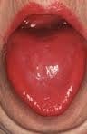
niacin deficiencyWhat Is A Fiery Red, Smooth, Burning Tongue?

A fiery red, smooth, burning tongue is an alteration in tongue tissue that is characteristic of advanced niacin deficiency.1

Q: How does niacin deficiency cause the tongue to be red and sore?

A: Niacin is an essential B vitamin that is required for a healthy tongue and by all body cells as well. Deficiency first shows in tissues with rapid cellular turnover, such as mucosal cells of the tongue.

When absorbed from the small intestines, niacin (the form in food) becomes the active form niacinamide. Niacinamide is converted by the body into co-enzymes which are present in all cells. These enzymes function in oxidation-reduction reactions essential for release of energy from carbohydrates, fats, and proteins and are needed as components for more than 200 enzymes involved in metabolism.

In addition to producing energy, niacinamide is essential for healthy skin and the mucosal lining of the digestive tract, normal functioning of the brain and nervous system, and production of steroid hormones from adrenal glands and hormones from sex glands.

What Is A Fiery Red, Smooth, Burning Tongue In Celiac Disease and/or Gluten Sensitivity?

Sources:
  1. Krause’s Food, Nutrition, & Diet Therapy. 10th Edition. Kathleen Mahan, Sylvia Escott-Stump. 2000. W.B. Saunders Company. []

Vitiligo

Areas of the Pharynx Where Cancer May Occur. Courtesy Wikimedia.

What Is Cancer Of The Pharynx?

C ancer of the pharynx is a malignant growth of stratified squamous cells that line the pharynx, or throat.

Q: What are stratified squamous cells that line the pharynx?

A: Stratified squamous cells are thin, flat epithelial cells consisting of several layers forming the surface mucosa that protects underlying layers of the pharynx. These cells lie very close together and have no blood vessels.

The pharynx is the upper passageway of the throat where swallowing food and fluids from the mouth first pass on the way to the esophagus and breathing air passes from the nose and mouth to and from the lungs.

The pharynx provides for speech production, adequate taste reception, and proper hearing through equalization of air pressure in the eustachian tubes.

The pharynx has three areas and pharyngeal cancer can occur in any of them: the nasopharynx behind the nose, the oropharynx behind the mouth, and the hypopharynx or laryngopharynx, just above the larynx. Tonsils, adenoids, and other lymph tissue lie at the back of the throat.

Unfortunately, cancers of the pharynx tend not to be detected until late in their course when symptoms become evident. This makes the prognosis poor. See symptom below.

What Is Cancer Of The Pharynx In Celiac Disease and/or Gluten Sensitivity?

Scleroderma (Systemic Sclerosis)

Geographic Tongue Due to Riboflavin Deficiency.

 What Is Geographic Tongue?

G eographic tongue is a painful alteration in tongue tissue that is characteristic of riboflavin (vitamin B2) deficiency.1

Q: How does the tongue surface appear?

A: Areas of inflamed tissue with flattened papillae develop among areas of normal tissue, giving a map-like appearance to the tongue surface. There are often swollen papillae that hurt or sting, such as is seen in the photo to the left.

A diet that does not provide adequate amounts of riboflavin and any condition that 1) interferes with riboflavin absorption from the gut, 2) depletes the vitamin from the body, 3) overuses the vitamin in the body, or 4) adversely affects its use in the body will result in riboflavin deficiency causing geographic tongue.

Riboflavin is essential for converting foodstuffs into energy, maintaining the normal lifespan of red blood cells and maintaining healthy skin, eyes, hair, and nervous system. Riboflavin is converted into its active forms, flavin adenine dinucleotide (FAD) and flavin mononucleotide (FMN). These enzymes are primarily involved as co-factors in oxidation reduction reactions that are essential for cellular energy production and respiration.

Riboflavin is required for the conversion of pyridoxine (vitamin B6) to its functional form and the production of niacin (vitamin B3) from the amino acid tryptophan. Riboflavin has a role in antioxidant status by activating glutathione reductase, which regenerates used glutathione, a powerful antioxidant.

Respiratory infection, exercise, certain diseases, drugs and hormones can adversely influence riboflavin status.2 Stress is a major depleter of riboflavin.

What Is Geographic Tongue In Celiac Disease and/or Gluten Sensitivity?

Sources:
  1. Krause’s Food, Nutrition, & Diet Therapy. 10th Edition. Kathleen Mahan, Sylvia Escott-Stump. 2000. W.B. Saunders Company. []
  2. Lakshmi AV. Riboflavin metabolism–relevance to human nutrition. Indian J Med Res. 1998 Nov;108:182-90. []

Anti-Endomysium Antibodies (EMA) Present In Blood

Smooth muscle. Courtesy pathguy.com
Smooth Muscle. Courtesy pathguy.com

What Are Anti-Endomysium Antibodies?

Anti-endomysium antibodies (EmA) are connective tissue autoantibodies produced in persons who have inherited the genes for celiac disease, an autoimmune disease, and are reacting to gluten in their diet.

Autoantibodies are abnormal in that they attack the body’s own tissue.

Q: What is endomysium?

A: Endomysium is the delicate connective tissue that surrounds individual muscle fibers. The autoantigen, or target, that stimulates the autoimmune response is the naturally occuring enzyme in endomysium called tissue transglutaminase (tTG), or more specifically tranglutaminase-2 (TG2). Anti-tissue transglutaminase antibodies recognize the same antigen as EmA, from which they differ in terms of detection method.

Anti-endomysium antibodies (EmA) are tested by the indirect immunofluorescence method and directed against “reticulin-like” fibres in connective tissue around smooth muscle fibres in the esophagus, liver, stomach, and bladder of monkeys, in the sections of the jejunum and kidneys of rats and in sections of the human umbilical cord. In comparison, for the determination of anti-tissue transglutaminase IgA and IgG antibodies, ELISA with human extractive or recombinant transglutaminase is recommended.1

EmA‐binding patterns in serum samples from patients with celiac disease have proved to be exclusively TG2‐targeted and the correlation between EmA and TG2 antibodies is therefore good. Evidence shows that celiac autoantibodies are produced in the small‐bowel mucosa.2

What Are Anti-Endomysium Antibodies In Celiac Disease and/or Gluten Sensitivity?

Sources:
  1. Trigoni E, Tsirogianni A, Pipi E, Mantzaris G, Papasteriades C. Celiac disease in adult patients: specific autoantibodies in the diagnosis, monitoring, and screening. Autoimmune Dis. 2014;2014:623514. doi: 10.1155/2014/623514. []
  2. Salmi TT, Collin P, Korponay-Szabó IR, Laurila K, Partanen J, Huhtala H, Király R, Lorand L, Reunala T, Mäki M, Kaukinen K. Endomysial antibody‐negative coeliac disease: clinical characteristics and intestinal autoantibody deposits.Gut. 2006 Dec;55(12):1746-53. []

Anti-tissue Transglutaminase Antibodies (tTG) Present In Blood

Image depicts tissue transglutaminase (tTg).
Image Depicts Tissue Transglutaminase (tTG).

What Are Anti-tissue Transglutaminase Antibodies?

Anti-tissue transglutaminase antibodies (anti-tTG) are connective tissue autoantibodies and can be detected in blood samples from affected persons who are reacting to gluten in the diet.

Autoantibodies are abnormal because they attack the body’s own tissue, which in the case of these antibodies is tissue transglutaminase 2 (TG2).

Q: What is tissue transglutaminase 2 (TG2)?

A: Tissue transglutaminase 2 (TG2) is an enzyme that appears in many cell locations and is particularly abundant in endothelial cells that line the small intestine. It has been implicated in a variety of cellular processes, such as differentiation, cell death, inflammation, cell migration and wound healing.

The cell appears to adapt the dynamics of this enzyme to meet specific sub-cellular needs or to respond to stress or other stimuli. Substantial evidence indicates that the location of TG2 within cells is critical for the regulation of its various biochemical activities, which subsequently trigger diverse downstream events,1

Although initially studied as an enzyme within cells, TG2 is now known to be secreted also into the extracellular space (between cells) or onto the cell surface.1

Abnormal activation of TG2 or deregulation of its function(s) is involved in a variety of human diseases, such as celiac disease, diabetes, neurodegenerative diseases, multiple sclerosis and rheumatoid arthritis. A role in inflammatory disorders and septic shock has also been shown. Moreover, multiple studies have revealed elevated TG2 expression in many types of cancer cells.1

What Are Anti-tissue Transglutaminase Antibodies In Celiac Disease and/or Gluten Sensitivity?

Sources:
  1. Piacentini M, D’Eletto M, Farrace MG, Rodolfo C, Del Nonno F, Ippolito G, Falasca L. Characterization of distinct sub-cellular location of transglutaminase type II: changes in intracellular distribution in physiological and pathological states. Cell Tissue Res. 2014 Dec;358(3):793-805. doi: 10.1007/s00441-014-1990-x. [] [] []

Autoimmune Polyglandular Syndromes 

Zincemia low zinc level symptom of celiac disease and glutenWhat Is Zincemia?

Z incemia means the zinc level in blood plasma is too low to meet metabolic needs of the body for this mineral.

Q: How important is a normal blood level of zinc?

A: A low blood level of zinc is characterized by widespread alterations in energy metabolism, growth, hemoglobin, carbon dioxide transport, hormone activity, insulin storage, many enzyme activities, prostaglandin function, collagen production, male fertility, protein synthesis, and vitamin A metabolism.

What Is Zincemia In Celiac Disease and/or Gluten Sensitivity?

Juvenile Diabetes (Type 1 Diabetes Mellitus)

Muscle pain in celiac disease and gluten sensitivityWhat Is Chronic Muscle Pain And Tenderness?

M uscle pain and tenderness, also called myalgia, is a disorder of muscle tissue.

Muscle pain that is ongoing or chronic can result from a variety of disorders, infections, overuse, injury, as an unwanted effect of certain medications, toxic substances, celiac disease, and certain nutritional deficiencies.

What Is Muscle Pain and Tenderness In Celiac Disease and/or Gluten Sensitivity?

Chronic Bullous Dermatosis of Childhood or Linear IgA Dermatosis

Muscle spasm and cramping in celiac disease and gluten sensitivity symptomWhat Are Muscle Spasm, Muscle Cramps?

M uscle spasm and muscle cramps are  disorders of muscle function caused by painful involuntary contractions of affected skeletal muscles characterized by limited movement.

Q: What is the difference between muscle spasm and cramps?

A: Cramps are stronger and more painful than spasms, occurring while the muscle is in its most shortened state.

Skeletal muscles are those that move the body skeleton to do work as we choose. Examples are muscles that move the mouth, arms,  hands, legs, and feet.

What Are Muscle Spasm, Muscle Cramps In Celiac Disease and/or Gluten Sensitivity?

Dermatitis Herpetiformis In Childhood

muscle wastingWhat Is Muscle Wasting?

M uscle wasting, or atrophy, is a muscle disorder resulting from the loss of muscle tissue which is characterized by thin muscles that have reduced strength and endurance.

What Is Muscle Wasting In Celiac Disease and/or Gluten Sensitivity?