Low stomach acid is common in celiac disease and dermatitis herpetiformis. It is also common in the general population, as well, affecting 50% of people age 60 years and about 80% by age 85 years. Nevertheless, low stomach acid is not generally looked for as a cause of acute and chronic disorders that rob health with far-reaching effects.
Is Low Stomach Acid New?
No. Low stomach acid (hypochlorhydria), has been well investigated much of the past century in both the general population and those with gluten sensitivity. For example, a 1985 study investigating gastric acid secretion in 116 subjects with dermatitis herpetiformis found that 41% had low stomach acid and 26% were achlorhydric (no acid). Of those older than 50 years, 47% were achlorhydric. When compared to subjects with celiac disease, the frequency of achlorhydria was significantly higher in those with dermatitis herpetiformis than in those with coeliac disease. There was no correlation between achlorhydria and small intestinal villous atrophy (damage).
Why Is Low Stomach Acid Overlooked?
Failure to understand nutrition and malabsorption…an area of science that is barely taught in medical schools is a big factor. Also,
Do you know someone who bruises easily? Do the marks develop into dark bluish swellings that hurt and take a long time to go away? If you have celiac disease, perhaps you had this problem before starting the gluten-free diet.
Easy bruising is a type of ecchymosis, or superficial bleeding under the skin or mucous membrane, common in untreated celiacs – and may be the only symptom of celiac disease. While always a sign of an underlying problem,
Close-up Slice of a Small Intestinal Villus Showing How Enterocytes Appear Tightly Lining the Entire Outside Surface Of A Villus. Courtesy Cleo Libonati
What Is Increased Intestinal Permeability?
Increased intestinal permeability is characterized by dysfunctional intestinal permeability (leakiness) allowing for the penetration of harmful entities from the gut into the bloodstream such as undigested proteins and microbes. The popular name is “leaky gut.”
Q: Why does intestinal permeability increase?
A: Intestinal permeability is an essential function of the small intestinal mucosal lining by which wanted substances such as properly digested foodstuffs are allowed to permeate through the lining to enter the body via the bloodstream and lymphatics. At the same time unwanted substances are kept out.
The mucosal lining is one cell thick and makes up the surface between the digested foodstuffs inside the hollow of the intestine and the underlying tissues.
The mucosal lining is covered by millions of microscopic finger-like structures called villi that project toward the inside of the intestine giving the appearance of a shag rug.
Each one, called a villus, contains a capillary bringing blood to absorb nutrients, a vein to take away nutrients, and a lacteal to absorb and take away digested fat. Its wall is made up of a single layer of tightly connecting cells, called enterocytes.
This single layer of cells separates the contents of our small intestine from the lamina propria (underlying tissues of the small intestine) and the rest of our body. Breaching of this single layer of cells by leakiness can expose lymphocytes (immune cells) located in the lamina propria to a myriad of microorganisms and food antigens, leading to immune reactions.1
To protect the body from unwanted substances, a gatekeeping barrier system operates to regulate the passage of nutrients, or permeation, through the surface mucosal lining. This system acts to seal the inside body from the gut.
The integrity of intestinal permeability is determined by interactions among several barrier components including the unstirred water layer, mucosal surface hydrophobicity, the surface mucous coat, and cell factors (especially tight junctions).
Tight junctions hold cells tightly together side-by-side to prevent unwanted substances from passing through the lining. Tight junctions are complex structures comprising over 50 proteins, such as the claudin proteins which are considered to be the structural backbone of tight junctions.
Tight junctions include a series of special proteins forming fibrils (springy like proteins) that cross the plasma membrane and interact with proteins in the adjoining cells. Tight junctions are regulated by the protein zonulin.2
If zonulin deregulates from the action of substances such as gliadin (gluten in wheat) and bacteria, the tight junction barrier fails which results in increased intestinal permeability. Dysfunction of the barrier system allows unwanted substances to enter the body where they are damaging to many tissues.
Tight junction dysfunction has been shown to be a part of certain autoimmune diseases such as celiac disease, type I diabetes mellitus, multiple sclerosis, and rheumatoid arthritis. Other diseases associated are cancer, allergies, and infections.3
Important gastrointestinal infections that cause leaky gut include rotavirus, parasites, pathogenic bacteria (escherichia coli, clostridium difficile), and mycotoxins produced by fungi found in stored grain and dried fruit.4
Fortunately, the presence of some commensal (friendly intestinal bacteria) and probiotic strains leads to an increase in tight junctions proteins at the cell boundaries and in some cases prevents or reverses the adverse effects of pathogens, food and stress. Various dietary components are also known to regulate epithelial permeability by modifying expression and localization of tight junctions proteins.2
What Is Increased Intestinal Permeability In Celiac Disease and/or Gluten Sensitivity?
Sources:
Fahardi A, Banan A, Fields J, Keshavarzian A. Intestinal barrier: an interface between health and disease. Journal of Gastroenterology and Hepatology. 2003; 18: 479-497. [↩]
Ulluwishewa D, Anderson RC, McNabb WC, Moughan PJ, Wells JM, Roy NC. Regulation of tight junction permeability by intestinal bacteria and dietary components. J Nutr. 2011 May;141(5):769-76. doi: 10.3945/jn.110.135657. [↩] [↩]
Fasano A. Zonulin and Its Regulation of Intestinal Barrier Function: The Biological Door to Inflammation, Autoimmunity, and Cancer. Physiological Reviews. January 2011Vol. 91no. 151-175DOI: 10.1152/physrev.00003.2008 [↩]
Farhadi A, Banan A, Fields J, Keshavarzian A. Intestinal barrier: an interface between health and disease. Journal of Gastroenterology and Hepatology. 2003;18:479-91. [↩]
phenomena that can originate in the eye when the vitreous gel pulls on or separates from the retina such as during or after a posterior vitreous detachment or from the visual centers of the brain…
Small clumps of cells or tissue that “float” in the clear gel of the vitreous body, the large chamber of the eyeball behind the lens and before the retina. Light travels through the clear lens…
Calcium is the most abundant mineral in the body. About 99% of this essential nutrient is contained in bones and teeth with the rest being in blood and other tissues. Calcium is needed for strong bones and teeth and for nerve conduction, muscle contraction, heart muscle function, blood pressure regulation, glycogen to glucose conversion, initiation of blood clotting, many hormone actions, many enzyme activities and making acetylcholine, an important chemical for nerve transmission. Calcium plays a part in the prevention of colon cancer.
Most importantly, calcium opposes phosphorus as a buffer to maintain the acid-alkaline balance of the blood and is critical for milk production in the nursing of infants.
Calcium absorption in the small intestine is complex and has specific requirements.
Editor’s note: In this case report of infants with severe malabsorption from celiac disease, the treating physicians found copper deficiencies based on blood studies that showed severe low copper levels and white blood cell count. Treatment required copper supplementation in addition to the gluten-free diet. Normally, in the last few months of gestation, an infant stores a large amount of copper in their liver. This storage must last about 6 months because infants must derive their nourishment from copper-poor milk. This case report shows dramatically the terrible effect of malabsorption coupled with a naturally occurring huge demand for copper that could not be satisfied through digestion.
Osteoporosis, or brittle bones, is a generalized bone disorder involving the slow loss of bone mass throughout the skeleton that results in diminished bone mineral density (BMD). Thinning, fragile bones maintain normal cell appearance but have a rapid turnover so that more bone is taken up and removed than is laid down. The result is bone weakness that predisposes people with osteoporosis to fractures.
Osteopenia refers to the progression of bone tissue loss in the range between normal to osteoporosis.
What are Bones?
Bones are dynamic structures made up of living connective tissue and certain minerals. Connective tissue provides the shape of bones and holds calcium phosphate mineral for hardness and
Editor’s note: The study below, investigating whether the degree of villous atrophy (intestinal damage) correlates with the symptoms that are presented, found they do not. Therefore, more research is needed to find out why symptoms do not correlate with the degree of intestinal damage.
The pathologic range of villous atrophy seen on small intestinal biopsies ranges from severe (total villous atrophy and subtotal villous atrophy) to milder, partial villous atrophy.
Cancel your subscription?
We're sorry to see you go.
Please select a reason for cancellation.
Please provide additional details.
Your subscription is cancelled
You won't be billed again. If you change your mind, you can subscribe again at any time.
Cancelled
Cancellation request submitted
We've received your request. Your subscription remains active while we process it. We'll email you once it's confirmed.
Cancellation pending
Pause subscription
Your subscription payments will be paused. Your existing access will remain active while your subscription is paused.
Please choose a valid future date.
Resume subscription
Your subscription payments will resume on the normal billing schedule.
Access Content
Affiliate Terms
By joining our Affiliate Program, you agree to the following terms:
1. Eligibility: You must be 18 years or older to participate. We reserve the right to refuse service to anyone for any reason at any time.
2. Promotional Guidelines: Affiliates must not engage in misleading advertising or spam. Promotions must accurately represent our brand and products.
3. Affiliate Links: You must use your unique tracking link to receive credit for referred sales.
4. Prohibited Use: You may not use our branding in a way that may confuse customers or misrepresent your relationship with us.
5. Termination: We reserve the right to terminate your account at any time for breach of these terms.
6. Changes to Agreement: We may update these terms at any time. Continued participation implies acceptance of any changes.
By continuing, you agree to the terms of this Affiliate Agreement.


 Low stomach acid is common in celiac disease and dermatitis herpetiformis. It is also common in the general population, as well, affecting 50% of people age 60 years and about 80% by age 85 years. Nevertheless, low stomach acid is not generally looked for as a cause of acute and chronic disorders that rob health with far-reaching effects.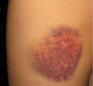
 Do you know someone who bruises easily? Do the marks develop into dark bluish swellings that hurt and take a long time to go away? If you have celiac disease, perhaps you had this problem before starting the gluten-free diet.
 Risotto is a unique rice dish with a full-bodied creamy mouth feel. Try it once and you are sure to make it often.
 Calcium is the most abundant mineral in the body. About 99% of this essential nutrient is contained in bones and teeth with the rest being in blood and other tissues. Calcium is needed for strong bones and teeth and for nerve conduction, muscle contraction, heart muscle function, blood pressure regulation, glycogen to glucose conversion, initiation of blood clotting, many hormone actions, many enzyme activities and making acetylcholine, an important chemical for nerve transmission. Calcium plays a part in the prevention of colon cancer.
 Editor’s note: In this case report of infants with severe malabsorption from celiac disease, the treating physicians found copper deficiencies based on blood studies that showed severe low copper levels and white blood cell count. Treatment required copper supplementation in addition to the gluten-free diet. Normally, in the last few months of gestation, an infant stores a large amount of copper in their liver. This storage must last about 6 months because infants must derive their nourishment from copper-poor milk. This case report shows dramatically the terrible effect of malabsorption coupled with a naturally occurring huge demand for copper that could not be satisfied through digestion.
 Osteoporosis, or brittle bones, is a generalized bone disorder involving the slow loss of bone mass throughout the skeleton that results in diminished bone mineral density (BMD). Thinning, fragile bones maintain normal cell appearance but have a rapid turnover so that more bone is taken up and removed than is laid down. The result is bone weakness that predisposes people with osteoporosis to fractures.
 Editor’s note: The study below, investigating whether the degree of villous atrophy (intestinal damage) correlates with the symptoms that are presented, found they do not. Therefore, more research is needed to find out why symptoms do not correlate with the degree of intestinal damage.