Parathyroid Glands in the Neck. Courtesy Wikipedia.com
What Is Secondary Hyperparathyroidism?
S econdary hyperparathyroidism is a parathyroid disorder resulting from hypocalcemia (low blood calcium level) that is characterized by excessive production of parathyroid hormone in the attempt to normalize the low blood calcium by releasing calcium from bone.
Parathyroid hormone is produced by the four pea sized parathyroid glands that are located on the thyroid gland in the front of the neck. In part, because the thyroid and parathyroid glands share the same anatomic place in the body and because they have similar names, they are often confused although they have completely different actions.
Parathyroid hormone regulates calcium and the opposing mineral phosphorus in the blood. In secondary hyperparathyroidism, calcium blood levels are low to normal while phosphorus levels are increased which stimulates the outpouring of parathyroid hormone.
Q: How does secondary hyperparathyroidism differ from primary hyperparathyroidism?
A: In primary hyperparathyroidism blood calcium is high and phosphorus is low, which is the opposite of secondary hyperparathyroidism.
The most common cause of secondary hyperparathyroidism is kidney disease causing failure to reabsorb calcium followed by vitamin D deficiency and malabsorption.
What Is Secondary Hyperparathyroidism In Celiac Disease and/or Gluten Sensitivity?
Glands which empty their hormone substances into the bloodsteam and from there to a targeted part of the body such as insulin produced by the pancreas and thyroxine produced by the thyroid gland. They are…
This 21 year-old woman (right) appears as old as her 70 year-old grandmother (left). Courtesy Prof Dr Chua Chung Nen
What Is Acquired Cutis Laxa?
A cquired cutis laxa is an uncommon skin disorder characterized by abnormal reduction and degeneration of elastic fibers of the skin that can appear simply as thick, saggy skin with loose folds to severe involvement showing a premature aged appearance.
Q: What are elastic fibers of the skin?
A: Elastic fibers of the skin are connective tissue found in the dermis, which is the layer of skin under the epidermis, or surface layer. They hold the shape of skin and are important for wound healing in the development of scars.
What Is Cutis Laxa In Celiac Disease and/or Gluten Sensitivity?
What Is Vitiligo? V itiligo is a pigmentation disorder of the skin characterized by permanent loss of melanocytes in defined areas and, in some patients, antibodies to melanin. Vitiligo has significant psychological impact if occurring before…
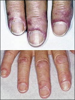
D ermatomyositis is a rare autoimmune systemic disease of the connective tissue that is characterized by inflammatory and debilitatingdegenerative changes in the muscles and in the skin.
Dermatomyositis results in symmetric, proximal muscle weakness of limbs (upper arms and legs), and skin manifestations. 50-70% of patients have circulating myositis-specific auto-antibodies.
The course of dermatomyositis is unpredictable being marked by spontaneous flare-ups and remissions. It can begin slowly or abruptly according to the factor that is triggering the onset such as infection, medications like phenytoin, and autoimmune disease.
Q: What are the skin manifestations of dermatomyositis?
A: Classic skin manifestations of dermatomyositis include these features:
The heliotrope rash (lilac color) on upper eyelids.
Rash on face, neck, shoulders, upper chest, elbows, knees, knuckles, and back.
Gottron’s papules (scaly, red eruptions or patches over the knuckles, elbows, and knees).
The V-sign (rash front of neck and chest).
The shawl sign (rash distribution on shoulders and back).1
Additional cutaneous manifestations are described below under symptoms.
Dermatomyositis is associated with an increased risk of cancer, other autoimmune diseases, such as lupus and psoriasis, and it can be a complication of interferon-α therapy.About 1 person in 100,000 are affected according to various studies. While it affects all ages, women have twice the occurence of men.
There is no cure for dermatomyositis, but the symptoms can be treated. Options include medication, physical therapy, exercise, heat therapy (including microwave and ultrasound), orthotics and assistive devices, and rest. The standard treatment for dermatomyositis is a corticosteroid drug, given either in pill form or intravenously. Immunosuppressant drugs, such as azathioprine and methotrexate, may reduce inflammation in people who do not respond well to prednisone.2
What Is Dermatomyositis In Celiac Disease and/or Gluten Sensitivity?
Sources:
Marvi U, Chung L, Fiorentino DF. Clinical presentation and evaluation of dermatomyositis. Indian J Dermatol. 2012 Sep;57(5):375-81. doi: 10.4103/0019-5154.100486. [↩]
National Institute of Neurological Disorders and Stroke. [↩]
Swollen Tongue Causing Tooth Indentations. Notice the Accompanying Denuded Area Due to Riboflavin Deficiency and Mild Candida Overgrowth. GFW
What Is A Pale, Smooth, Burning Tongue?
A pale, smooth, burning tongue is an alteration in tongue tissue characteristic of iron deficiency. The tongue is also swollen.1
Additionally, the sore tongue surface may be invaded by candida yeast which takes advantage of the sore tissue.
Iron deficiency itself increases susceptibility to infection.
Q: What is iron deficiency?
A: Iron deficiency results when the level within cells is too low to meet metabolic needs of the body for this mineral.
Deficiency is characterized by impaired red blood cell formation, free-radical disposal, oxygenation of cells, immune response to infection, enzyme activity, cognitive performance, digestion, nail structure, and fetal health.2
Iron is an essential mineral that is required for normal body function. Almost two-thirds of iron in the body is found in hemoglobin, the protein in red blood cells that carries oxygen to tissues. Smaller amounts of iron are found in myoglobin, a protein that helps supply oxygen to muscle, and in enzymes that assist biochemical reactions.
Iron is also found in proteins that store iron for future needs and that transport iron in blood. Iron stores are regulated by intestinal iron absorption.3
What Is A Pale, Smooth, Burning Tongue In Celiac Disease and/or Gluten Sensitivity?
Sucrose intolerance is the inability to digest sucrose, a widely available sugar, while sucrosemia is the abnormal presence of sucrose in the bloodstream.
Q: Why cannot the body digest sucrose?
A: Sucrose, such as cane or beet sugar, is a double molecule sugar which must first be digested before being absorbed from the gut into the bloodstream. That is, sucrose must be split into its component single molecules of fructose and glucose, which are then properly absorbed.
The inability to properly digest sucrose results directly from low production and activity of sucrase in the small intestine. Sucrase is the specific enzyme that splits or digests sucrose.
Undigested sucrose does not remain idle. Its presence acts osmotically to draw water from the body into the intestine, causing watery diarrhea.
Meanwhile, microbiota (normal bacteria) in the colon eagerly ferment the abnormally present sucrose that arrives from the small intestine. Fermentation generates short-chain fatty acids and hydrogen gas, which results in bloating pain.1
In sucrosemia, sucrose molecules abnormaly pass through an unhealthy small intestinal lining and enter the bloodstream where there presence is abnormal. Sucrose in the blood is filtered out by the kidneys and excreted in urine.
Positive response to a breath hydrogen test (BHT), involving 1 – 3 hours of time post ingestion of sucrose test dose, signifies malabsorption in the small intestine and fermentation in the colon. If BHT is positive before 60 minutes, the result implies bacteria is abnormally present in the small intestine, causing fermentation there. Endoscopy is used to measure sucrase activity in tissue samples.
What Is Sucrose Intolerance And Sucrosemia In Celiac Disease and/or Gluten Sensitivity?
Swollen Tongue Causing Tooth Indentations. Notice the Accompanying Denuded Area Due to Riboflavin Deficiency and Mild Candida Overgrowth. GFW
What Is A Pale, Smooth, Burning Tongue?
A pale, smooth, burning tongue is an alteration in tongue tissue characteristic of iron deficiency. The tongue is also swollen.1
Additionally, the sore tongue surface may be invaded by candida yeast which takes advantage of the sore tissue.
Iron deficiency itself increases susceptibility to infection.
Q: What is iron deficiency?
A: Iron deficiency results when the level within cells is too low to meet metabolic needs of the body for this mineral.
Deficiency is characterized by impaired red blood cell formation, free-radical disposal, oxygenation of cells, immune response to infection, enzyme activity, cognitive performance, digestion, nail structure, and fetal health.2
Iron is an essential mineral that is required for normal body function. Almost two-thirds of iron in the body is found in hemoglobin, the protein in red blood cells that carries oxygen to tissues. Smaller amounts of iron are found in myoglobin, a protein that helps supply oxygen to muscle, and in enzymes that assist biochemical reactions.
Iron is also found in proteins that store iron for future needs and that transport iron in blood. Iron stores are regulated by intestinal iron absorption.3
What Is A Pale, Smooth, Burning Tongue In Celiac Disease and/or Gluten Sensitivity?
Microscopic Image Showing a Pink Collagen Band in Collagenous Colitis.
What Is Collagenous Colitis?
C ollagenous colitis is a disease of the large intestine (colon) that is characterized by microscopic inflammation of the surface mucosal lining and an abnormally thickened collagen band of tissue that develops wthin the lining of the colon.
The thicker than normal layer of collagen of at least 10 µm (reference value: 2–7 µm) can vary in different locations. Inflammation occurs with increased numbers of lymphocytes (white blood cells) and plasma cells and epithelial (surface cell) damage. These changes can only be seen under microscopic examination of multiple biopsied tissue samples taken during a colonoscopy procedure.
Q: What is collagen?
A:Collagen is a strong, fibrous protein found in connective tissue of the colon and many other tissues such as tendons. The normal basement membrane in the bowel consists mainly of collagen type IV, laminin, and fibronectin. The increased collagen band observed in collagenous colitis consists basically of collagen type I and III, which are the subtypes produced by repair functions, indicating a reactive origin to some irritant or drug.1
The biopsies should preferably be taken from the ascending colon, since the pathological hallmarks may be absent in the descending colon, and in the normally occurring thicker collagen layer in the rectosigmoid region.1 Inflammation of the ileum (last segment of the small intestine next to colon) is common.2
Endoscopy and radiological (x-ray) examinations are usually normal.3
Autoimmune disorders are frequently seen in adult patients with collagenous colitis.4 In the study below by Koskela et al. concomittent autoimmune diseases were present in 53% of patients with collagenous colitis.5
Importantly, the finding of collagenous colitis in patients with autoimmune diseases may reflect the treatment with NSAIDs (non-steroidal anti-inflammatory drugs), such as Ibuprofin and aspirin, PPIs (proton pump inhibitors), and other drugs. However, if secondary forms of collagenous colitis are not taken into consideration, underlying, treatable diseases may be overlooked, while only the gastrointestinal symptoms are treated symptomatically or with budesonide (a steroid).6
Treatment with budesonide steroid is efficacious irrespective of bile acid malabsorption.7
Budesonide at a mean dose of 4.5 mg/day maintained clinical remission for at least 1 year in the majority of patients with collagenous colitis and preserved health-related quality of life without safety concerns. Treatment extension with low-dose budesonide beyond 1 year may be beneficial given the high relapse rate after budesonide discontinuation.8
See below for nutritional deficiency problems caused by steroid usage and steps to be taken for correction.
What Is Collagenous Colitis In Celiac Disease and/or Gluten Sensitivity?
Sources:
Ohlsson B. New insights and challenges in microscopic colitis. Therap Adv Gastroenterol. 2015 Jan;8(1):37-47. doi: 10.1177/1756283X14550134. [↩] [↩]
Bjørnbak C, Engel PJ, Nielsen PL, Munck LK. Microscopic colitis: clinical findings, topography and persistence of histopathological subgroups. Aliment Pharmacol Ther. 2011 Nov;34(10):1225-34. doi: 10.1111/j.1365-2036.2011.04865.x. [↩]
Abdo AA, Urbanski SJ, Beck PL. Lymphotcytic and collagenous colitis: the emerging entity of microscopic colitis. An update on pathophysiology, diagnosis and management. Canadian Journal of Gastroenterology. Jul 2003;17(7):425-32. [↩]
Leung ST, Chandan VS, Murray JA, Wu TT. Collagenous gastritis: histopathologic features and association with other gastrointestinal diseases. Am J Surg Pathol. 2009 May;33(5):788-98. doi: 10.1097/PAS.0b013e318196a67f. [↩]
Koskela RM, Niemela SE, Karttunen TJ, Lehtola JK. Clinical characteristics of collagenous and lymphocytic colitis. Scandanavian Journal of Gastroenterology. Sep 2004;39(9):837-45. [↩]
Ohlsson B. New insights and challenges in microscopic colitis. Therap Adv Gastroenterol. 2015 Jan;8(1):37-47. doi: 10.1177/1756283X14550134. [↩]
Bjørnbak C, Engel PJ, Nielsen PL, Munck LK. Microscopic colitis: clinical findings, topography and persistence of histopathological subgroups. Aliment Pharmacol Ther. 2011 Nov;34(10):1225-34. doi: 10.1111/j.1365-2036.2011.04865.x. [↩]
Münch A, Bohr J, Miehlke S, et al. Low-dose budesonide for maintenance of clinical remission in collagenous colitis: a randomised, placebo-controlled, 12-month trial. Gut. 2014 Nov 25. pii: gutjnl-2014-308363. doi: 10.1136/gutjnl-2014-308363. [↩]
Cancel your subscription?
We're sorry to see you go.
Please select a reason for cancellation.
Please provide additional details.
Your subscription is cancelled
You won't be billed again. If you change your mind, you can subscribe again at any time.
Cancelled
Cancellation request submitted
We've received your request. Your subscription remains active while we process it. We'll email you once it's confirmed.
Cancellation pending
Pause subscription
Your subscription payments will be paused. Your existing access will remain active while your subscription is paused.
Please choose a valid future date.
Resume subscription
Your subscription payments will resume on the normal billing schedule.
Access Content
Affiliate Terms
By joining our Affiliate Program, you agree to the following terms:
1. Eligibility: You must be 18 years or older to participate. We reserve the right to refuse service to anyone for any reason at any time.
2. Promotional Guidelines: Affiliates must not engage in misleading advertising or spam. Promotions must accurately represent our brand and products.
3. Affiliate Links: You must use your unique tracking link to receive credit for referred sales.
4. Prohibited Use: You may not use our branding in a way that may confuse customers or misrepresent your relationship with us.
5. Termination: We reserve the right to terminate your account at any time for breach of these terms.
6. Changes to Agreement: We may update these terms at any time. Continued participation implies acceptance of any changes.
By continuing, you agree to the terms of this Affiliate Agreement.