Contents
What Is Systemic Lupus Erythematosus?
[dropcap]S[/dropcap]ystemic lupus erythematosus (SLE) is a chronic autoimmune inflammatory disease that is characterized by involvement of multiple organs due to the production of antibodies to components of the cell nucleus.1 SLE has an unpredictable course of acute flare-ups and remissions.
Severity depends on the extent of organs affected with skin and nail involvement, called discoid lupus, being the least serious and inflammmation of the kidney, called lupus nephritis, being the worst.
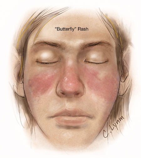
Nevetheless, a classic presentation is development of a rash over the cheeks and nose that resembles a butterfy with wings spread hence the name “butterfly rash.”
Symptoms are many and varied depending on the tissues affected and are often not specific, for example hair loss has a variety of causes. Symptoms can be confused by co-existence with other autoimmune disease such as Sjogren’s syndrome.
Systemic lupus erythematosus should be managed by a specialist. Symptoms can be controlled with steroid therapy, but this disease can be a cause of premature death mainly from active disease, organ failure (e.g., kidneys), infection, or cardiovascular disease from accelerated atherosclerosis.
Certain common medicines known to cause drug-induced lupus are:
- Isoniazid
- Hydralazine
- Procainamide
Other less common drugs may also cause the condition. These may include:
- Anti-seizure medications
- Capoten
- Chlorpromazine
- Etanercept
- Infliximab
- Methyldopa
- Minocycline
- Penicillamine
- Quinidine
- Sulfasalazine
Symptoms tend to occur after taking the drug for at least 3 to 6 months.2
Although there is a strong familial aggregation, the disease is relatively uncommon and most cases are sporadic.1 According to the Center for Diseases (CDC), lupus most commonly affects women of childbearing age but also occurs in infants, children, adolescents, and men with peak occurrence between ages 15 and 40. Blacks (and possibly Hispanics, Asians, and Native Americans) are affected more than Whites.
What Is Systemic Lupus Erythematosus In Celiac Disease and/or Gluten Sensitivity?
- Relationship between systemic lupus erythematosus and celiac disease. Systemic lupus erythematosus is an associated immune disorder in celiac disease. Reports give conflicting results regarding association with celiac disease. There is a high rate of false-positive serology results for celiac disease in SLE. In one study that examined 103 patients with SLE, none had either antiendomysial antibodies (EMA) or a mucosal lesion, but 23.3% had anti-gliadin antibodies. A study by Picceli et al. found that IgA-EmA signifying celiac disease was significantly associated with lupus with the presence of discoid lesions.3
- Relationship between systemic lupus erythematosus and gluten. The association between celiac disease and other immune disorders may be due to the sharing of a common genetic background, such as HLA antigens. However, in a very large study, involving 909 patients with celiac disease, Ventura and his associates found that the development of immune disorders in Celiac Disease was clearly related to the duration of exposure to gluten.4
- Relationship between systemic lupus erythematosus and autoimmunity. The association of celiac disease and SLE may shed light on the pathogenesis of autoimmune disorders in patients with celiac disease since anti-DNA antibodies from a patient with celiac disease induced experimental SLE in a mouse model.5
- Relationship between systemic lupus erythematosus and vitamin D. Vitamin D deficiency is associated with lupus and is a classic finding in celiac disease. Vitamin D is a prohormone that is required for normal immune system functioning. The physiologic and clinical consequences of vitamin D deficiency in systemic lupus erythematosus are not entirely known. Prospective studies of vitamin D in systemic lupus erythematosus are limited, but most cross-sectional studies show an inverse relationship between levels of vitamin D and disease activity. This suggests that normalizing of vitamin D may have benefits beyond bone health for patients with systemic lupus erythematosus.6
How Prevalent Is Systemic Lupus Erythematosus In Celiac Disease and/or Gluten Sensitivity?
- A 2012 Swedish study reported occurrence of systemic lupus erythematosus in 3.49% of patients with celiac disease.7
- A 2013 Brazilian study found a 5.5% occurrence of endomysium autoantibodies signifying celiac disease in patients with lupus having the presence of discoid lesions.3
- A study reported occurrence of systemic lupus erythematosus in 1.3% of patients with dermatitis herpetiformis.8
What Are The Symptoms Of Systemic Lupus Erythematosus?
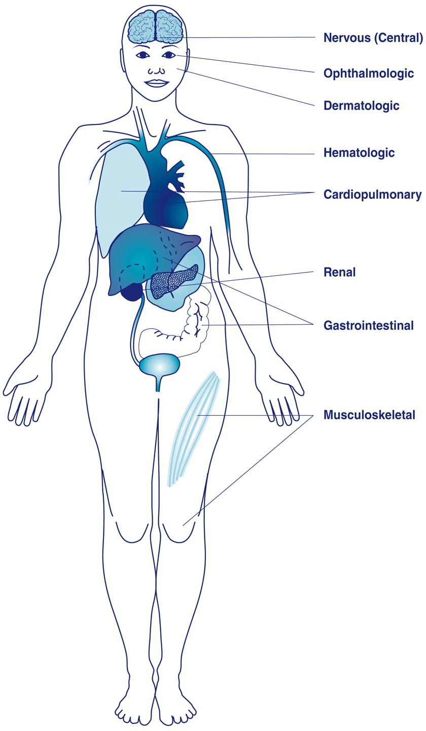
Systemic lupus erythematosus is usually marked by varied symptoms that tend to flare:
- Moderate to severe fatigue in most.
- Erythematous (red) “butterfly” rash over the nose and cheeks in many.
- Weight loss.
- Low grade fever under 101 dgrees Farenheit that may come and go with flares.
- Sensitivity to the sun in most.
- Ulcers in mucous membranes of mouth and throat in many.
- Hair loss in many.
- Mottling in fingers in many when exposed to cold.
- Depending on the type of involvement there can be raised, red, scaly discoid skin lesions, antinuclear antibodies, autoantibodies, pleuritis or pericarditis, arthritis (pain or swelling in joints), anemia, lymphopenia (low white blood cell count), and autoimmune thrombocytopenia which allows blood clot formations.
- Pain or irritation of the eyes in some because of inflammation of the uveal tract of the eye (called uveitis) caused by autoimmune antibodies produced in lupus.
- Calcinosis (calcium deposits in skin). Calcium deposits appear as hard bumps under the skin or in the muscle. Calcinosis most often occurs 1-3 years after the disease begins and is associated with damaged, inflamed, or necrotic skin.
- Possible stroke.
- Possible heart attack.
- Lower leg swelling, elevated blood pressure and possible kidney failure if kidneys become inflamed.
How Does Systemic Lupus Erythematosus Develop In Celiac Disease and/or Gluten Sensitivity?
- Systemic lupus erythematosuss results from an unclear autoimmune mechanism associated with celiac disease.
- Vitamin D deficiency is a factor in lupus, and in celiac disease it results from malabsorption. Most cross-sectional studies show an inverse relationship between levels of vitamin D and disease activity. That is, the better the vitamin D status in the body, the lower the inflammation activity of lupus.9
- A nationwide study in England investigating the reported association between vitamin D deficiency and the risk of developing immune-mediated diseases showed that patients with vitamin D deficiency have an increased rate of developing lupus.10
- Sunlight is a well known trigger for activating symptoms.
Does Systemic Lupus Erythematosus Respond To Gluten-Free Diet?
Studies are inadequate to determine effect of gluten free diet on the course of systemic lupus erythematosus, but case studies describe a beneficial effect.11,12
Vitamin D supplementation is advised to normalize blood levels which reduces disease activity.6
6 Steps To Improve Systemic Lupus Erythematosus In Celiac Disease and/or Gluten Sensitivity:
- [dropcap]1[/dropcap]Remove the Trigger. Maintain a Strict, Nutritious Gluten Free Diet:
[box type=”shadow” ]Treatment. This condition responds to the complete elimination of gluten, which is the required treatment that improves both systemic lupus erythematosus and gut health.
- Gut health is the foundation to restore ALL health. Restored health will enable you to maintain a strict gluten free diet, just as other life tasks will be easier.
- A strict gluten free diet means removing 100% of wheat, barley, rye and oats from the diet.
- Cutting out bread and other obvious sources of gluten is not good enough for recovery. Even 1/8th teaspoon of flour or bread crumb is enough to sustain the inflammation that is damaging your small intestine, causing increased permeability (leaky gut) and allowing undigested gluten to enter your body where it can damage structures and function, and instigate immune inflammatory responses.
Correct Your Individual Nutritional Needs.
- Eat foods that can replenish missing nutrients. Find them under NUTRIENT DEFICIENCIES.
- Take nutritional supplements as needed. Find them under NUTRIENT DEFICIENCIES.
Recovery. You should begin to feel better within a week and notice more energy as inflammation subsides and the absorbing cells that make up the surface lining of your small intestine are better able to function.
- Intestinal lining cells are replaced every 5 days. The healing process is like sunburn where the damaged surface layer of skin sloughs off and is replaced with new normal cells.
- Leaky gut normally resolves in two month after starting a gluten free diet and brings about a big improvement in health. Improvement in intestinal permeability precedes morphometric recovery (cell appearance and structure) of the small intestine in celiac disease.13
- The intestinal lining may take up to a year to heal.[/box]
- [dropcap]2[/dropcap] Reduce Inflammation. Foods to Eat and Foods Not to Eat:
Because gluten is inflammatory, eliminate OTHER inflammatory foods from your diet to reduce an additive effect to gluten. At the same time, try to eat foods that reduce inflammation (anti-inflammatory).
[box type=”shadow” ]Here Are Major Inflammatory Food Types That Reduce Healing:
- Damaging Foods. In susceptible persons, includes corn, dairy (cow), and soy. Lactose, the sugar in any animal milk disrupts intestinal permeability causing leaky gut.14
- Allergenic Foods. Includes foods that trigger the immune sytem to produce IgE antibodies. Allergy testing is the usual way to discover these offending foods.
- Shelf Stable Processed Foods. Includes any that contain additives and preservatives. Look for them on the nutrition label of the box or package. Additives and preservatives also disrupt intestinal permeability causing leaky gut.14
- Fats. Limit deep fried foods, trans-fats, saturated fats (animal fat/butter), and EXCESSIVE omega-6 fatty acid oils like corn oil. Rancid fats, sodium caprate (a medium chain fat), and sucrose monester fatty acid (a food grade surfactant) induce significant disruption of the intestinal barrier that causes leaky gut.14.
- Excessive Refined White Flours (bran layer removed). Includes products made from them such as cookies, bread, cakes, pies. Bran contains the vitamins and minerals that metabolize grains and slows the otherwise rapid entry of sugar from their digestion into the bloodstream. Also disrupt intestinal permeability causing leaky gut.14
- Refined Sugars. Includes white sugar, corn fructose and high fructose corn syrup.
- Certain Spices. Includes paprika and cayenne pepper which disrupt intestinal permeability causing leaky gut.14
- Alcohol and Caffeine. Disrupt intestinal permeability causing leaky gut.14[/box]
[box type=”shadow” ]Here Are Important Anti-Inflammatory Food Types to Promote Health:
- Fruits. Contain ample amounts of vitamins, minerals and phytochemicals which are naturally occuring components in plants that detoxify toxins, carcinogens (reducing the risk by 50%) and mutagens.
- Non-Starchy Vegetables. Support intestinal integrity and provide ample amounts of vitamins, minerals and phytochemicals. Includes green leafy vegetables such as lettuce and kale, also onion, broccoli, garlic, and others.
- High Quality Complex Carbohydrates. Provide vitamins, minerals, and fiber while boosting serotonin levels to help you relax and feel calm. Includes whole grains, legumes, and root vegetables such as carrots, parsnips, sweet potatoes, turnips, red beets, and others.
- Antioxidants. Protect the body from inflammatory oxidant molecules that continually occur and help us handle stress and reduce irritability. Includes vitamin C-containing foods such as lemon, grapefruit, apricot, Brussels sprouts and strawberries, and others. Also, includes vitamin E-containing foods such as nuts, seeds, avocado, olive oil, and others. Cocoa is good, too.
- Omega-3 Fatty Acids. Balance opposing omega-6 fatty acids and bad fats. Fish sources includes tuna, salmon, cod, and others. Plants sources include flax, chia seeds, canola oil, and others.
- Probiotics. Supply normal microbes needed for colon health and health of the body such as these fermented foods: yogurt, kefir, and unpasteurized apple cider vinegar.
- Prebiotics/ High Fiber Foods. Food with fiber keeps our population of colonic microbes healthy.
- Protective Herbs and Spices. See below #6 below for examples.[/box]
- [dropcap]3[/dropcap] Information Sheet You Can Take to Your Doctor or Other Health Professional:
Click here.
- [dropcap]4[/dropcap] Manage Your Medications Safely:
[box type=”shadow” ]
- Certain medications used to treat lupus deplete nutrients which requires supplementation to prevent nutritional deficiencies.
- Certain medications deplete vitamin D which worsens lupus and celiac disease.
Ask your doctor or pharmacist about this possible adverse effect if you are taking any of the drugs listed below. Do not stop prescribed medications without supervision.
This is not a complete listing.
1. Nutrient depleting drugs used to treat lupus include these:
ANTI-INFLAMMATORIES disrupt intestinal permeability which complicates celiac disease.
- Corticosteroids (Prednisone, Medrol®, Aristocort®, Decadron) deplete Calcium, Vitamin D, Magnesium, Zinc, Vitamin C, Vitamin B6, Vitamin B12, Folic Acid, Selenium, Chromium, Phosphorus.
- NSAIDS (Motrin®, Aleve®, Advil®, Anaprox®, Dolobid®, Feldene®, Naprosyn® and others) deplete Folic acid.
2. Common drugs that deplete vitamin D include these:
ANTACIDS / ULCER MEDICATIONS
- Pepcid®, Tagamet®, Zantac® deplete Vitamin D.
- Magnesium and Aluminum Antacid preparations (Gaviscon®, Maalox®, Mylanta®) deplete Vitamin D.
ANTI-INFLAMMATORIES disrupt intestinal permeability which complicates celiac disease.
- Corticosteroids (Prednisone, Medrol®, Aristocort®, Decadron) deplete Vitamin D.
ANTICONVULSANTS
- Phenobarbital and Barbituates; and Dilantin®, Tegretol®, Mysoline®, Depakane/Depacon® deplete Vitamin D.
BRONCHODILATORS
- Inhaled corticosteroid inhalers (Flovent, Pulmicort and others) that are breathed in on a daily basis as a long term therapy to reduce inflammation in airways deplete Vitamin D.
CHOLESTEROL DRUGS
- Colestid® and Questran® deplete Vitamin D.
LAXATIVES
- Metamucil, FiberCon, Citrucel, Colace, Glycolax, Milk of magnesia, Dulcolax deplete Vitamin D.
WEIGHT LOSS DRUGS THAT BIND FAT also interfere with absorption of some nutrients.
- Zenicol (Orlistat®) depletes Vitamin D.
[/box]
- [dropcap]5[/dropcap]Nutritional Supplements To Help Correct Deficiencies:
[box type=”shadow” ]
- Multivitamin/mineral combination that provides 100% once a day is useful to improve overall nutrient levels. This is a safe dose, but always check with your doctor to avoid interactions with medications.
- B-Vitamin Complex as prescribed to restore folic acid, vitamin B6, and vitamin B12 following blood test for status.
- Calcium citrate is the best absorbed of calcium supplements. Calcium carbonate is a poor choice.
- Vitamin D3 as prescribed following blood test for status.
- Chelated magnesium as prescribed, but do not take at same time as calcium because they compete for absorption.
- Zinc as prescribed following blood test for status.
- Chromium and/or selenium following blood test for status.
Storage Note: Store container tightly sealed, away from heat, moisture and direct light to avoid loss of potency. That is, in a safe kitchen cabinet – not in the bathroom or on the kitchen table.[/box]
- [dropcap]6[/dropcap]Manage Natural Remedies:
[box type=”shadow” ]Hydration:
- Eight glasses of water are recommended per day unless there is a contraindication such as kidney or heart disease. The Institute of Medicine recommends approximately 2.7 liters (91 ounces) of total water, from all beverages and foods, each day for women and 3.7 liters (125 ounces) daily of total water for men.
- If you are thirsty, drink water. Add fresh, squeezed lemon to water. Lemon is anti-inflammatory, alkalizing and provides vitamin C.
- Hydration Test: Urine should be pale yellow. Fingertips should be plump, without pruning but this may not be reliable when fingers are swollen with edema. Lips should be plump, without puckering. The feeling of thirst can be unreliable.
- What is wrong with soda, coffee, tea, and alcohol? These drinks are dehydrating, increase acid, and deplete nutrients.[/box]
[box type=”shadow” ]Carminatives. The following anti-inflammatory plant sources called carminitives help heal the digestive tract. They also tone the digestive muscles which improves peristalsis, thus aiding in the expulsion of gas from the stomach and intestine to relieve digestive colic and gastric discomfort.
Carminative Food Remedies:
- Raspberry.
- Carrot is also a cleansing digestive tonic.
- Grape is also bile stimulating and a cleansing remedy for sluggish digestion and laxative.
- Redbeets also stimulate and improve digestion and are easily digested.
- Cabbage also stimulates and improves digestion and is also a liver decongestant.
- Lettuce also stimulates and improves digestion and is also an alterative, meaning it improves the function of organs involved with the digestion and excretion of waste products to bring about a gradual change.
- Potatoes are antispasmodic (due to atropine like properties) and a liver remedy.
Carminative Herb Remedies:
- Sage is also a digestive, astringent, bile stimulant and energy tonic that heals the mucosa. Drink as tea or use in cooking.
- Chamomile, lemon balm, and fennel, (as a tea) also help relieve nervous tension.
- Parsley also relieves indigestion.
- Rosemary as a tea and in cooking also is a nervous system tonic for stress and fatigue, bile stimulant, and can relieve headaches and indigestion.
- Thyme is also soothing remedy useful for stimulating digestion of rich, fatty foods.
Carminative Spice Remedies:
- Cloves are also antispasmodic.
- Nutmeg is also useful for indigestion.
- Ginger.[/box]
[box type=”shadow” ]Exercise Helps:
Exercise improves circulation and rids the body of toxins but must be taken only to toleration because over exercise that stresses the body can provoke a flare-up of lupus symptoms.
- Walking is aerobic exercise that reconditions the whole body to improve stamina. Read more about Exercise and Fitness.
- Weight training builds muscle. Read more about Exercise and Fitness.
- Stretching improves flexibilty. Read more about Exercise and Fitness.
Note: Exercise is important, but the amount and type of exercise undertaken depends on your health. Your first priority is to heal. [/box]
What Do Medical Research Studies Tell About Systemic Lupus Erythematosus?
RESEARCH STUDY SUMMARIES
“Spectrum of autoantibodies for gastrointestinal autoimmune diseases in systemic lupus erythematosus patients.” This study investigating the occurrence of gastrointestinal organ-specific autoantibodies in 194 patients with systemic lupus and 103 healthy controls from Southern Brazil found that IgA-EmA (endomysium autoantibodies signifying celiac disease) was significantly associated with lupus with the presence of discoid lesions.
Anti-endomysium antibodies (IgA-EmA), anti-gastric parietal cells (GPC) antibodies, anti-smooth muscle antibodies (ASMA), anti-mitochondrial antibodies (AMA) and anti-LKM-1 (liver-kidney microsomal) were searched for using indirect immunofluorescence in the sera of patients and controls. The total positivity of antibodies in SLE patients was 14.4% (28/194) and differed significantly from healthy individuals (0.97%; p<0.001). IgA-EmA was more common in lupus patients than in controls (11/194; p=0.009), and one of these patients had dermatitis herpetiformis. Clinical association revealed that IgA-EmA was more common in SLE patients with discoid lesions. The frequency of anti-GPC (p=0.10), ASMA (p=0.16) and AMA (p=0.55) did not differ significantly between groups. No patient presented LKM-1 autoantibodies. One patient presenting anti-GPC was diagnosed with atrophic gastritis and pernicious anemia.15
“Hospital admissions for vitamin D related conditions and subsequent immune-mediated disease: record-linkage studies.” This public health study investigating a possible association between vitamin D deficiency and the risk of developing immune-mediated diseases found that patients with vitamin D deficiency may have an increased risk of developing some immune-mediated diseases, including Lupus although reverse causality or confounding cannot be ruled out.
A database of linked statistical records of hospital admissions and death registrations for the whole of England (from 1999 to 2011) were analyzed. Rate ratios for immune-mediated disease were determined, comparing vitamin D deficient cohorts (individuals admitted for vitamin D deficiency or markers of vitamin D deficiency) with comparison cohorts. After hospital admission for either vitamin D deficiency, osteomalacia or rickets, there were significantly elevated rates of Addison’sdisease, ankylosing spondylitis, autoimmune hemolytic anemia, chronic active hepatitis, celiac disease, Crohn’s disease, diabetes mellitus, pemphigoid, pernicious anemia, primary biliary cirrhosis, rheumatoid arthritis, Sjogren’s syndrome, systemic lupus erythematosus, thyrotoxicosis.16
“Increased risk of systemic lupus erythematosus in 29,000 patients with biopsy-verified celiac disease.” This nationwide study investigating a possible association between celiac disease (CD) and systemic lupus erythematosus (SLE) found that Individuals with CD were at a 3-fold increased risk of SLE compared to the general population. Although this excess risk remained more than 5 years after CD diagnosis, absolute risks were low.
The risk of SLE was compared in 29,048 individuals with biopsy-verified CD (villous atrophy, Marsh 3) from Sweden’s 28 pathology departments with that in 144,352 matched individuals from the general population identified through the Swedish Total Population Register. SLE was defined as having at least 2 records of SLE in the Swedish Patient Register. We used Cox regression to estimate hazard ratios (HR) for SLE.
During follow-up, 54 individuals with CD had an incident SLE. This corresponded to an HR of 3.49%. Beyond 5 years of follow-up, the HR for SLE was 2.54%.17
CASE REPORT SUMMARIES
“Systemic lupus erythematosus, celiac disease and antiphospholipid antibody syndrome: a rare association.” This case report decribes the beneficial treatment of a patient diagnosed with celiac disease who had systemic lupus erythematosus and antiphospholipid antibody syndrome, an association which has never been described before.18
“Celiac disease in systemic lupus erythematosus: a case report.” This case report describes the coexistence of systemic lupus erythematosus (SLE) with celiac disease. Systemic lupus erythematosus was diagnosed prior to celiac disease and initially SLE treatment was administered. After several years, when celiac disease symptoms developed, the diagnosis was corrected and additional treatment with a gluten-free diet was applied with beneficial effects.19
“Systemic lupus erythematosus with celiac disease: a report of five cases.” In five cases of SLE with villous atrophy on duodenal biopsy, only four had positive serological tests for celiac disease and only three had abdominal symptoms.20
“Diseases associated with dermatitis herpetiformis.” This study investigating the occurrence of associated diseases in a cohort of 305 patients with dermatitis herpetiformis (DH) who were followed for an average of 10 years compared with results from a cohort of patients with celiac disease demonstrated a prevalence of SLE in 1.3% of DH patients.8
Sources:
- http://www.cdc.gov/arthritis/basics/lupus.htm [↩] [↩]
- www.nlm.nih.gov/medlineplus/ency/article/000446.htm [↩]
- Picceli VF1, Skare TL, Nisihara R, Kotze L, Messias-Reason I, Utiyama SR. Spectrum of autoantibodies for gastrointestinal autoimmune diseases in systemic lupus erythematosus patients. Lupus. 2013 Oct;22(11):1150-5. [↩] [↩]
- La Villa G, Pantaleo P, Tarquini R, Cirami L, Perfetto F, Mancuso F, Laffi G. Multiple immune disorders in unrecognized celiac disease: a case report. World J Gastroenterol. 2003;9(6):1377-1380, Available at: http://www.wjgnet.com/1007-9327/9/1377.asp. [↩]
- Shamir R, Shoenfeld Y, Blank M, et al. The prevalence of coeliac disease antibodies in patients with the antiphospholipid syndrome. Lupus. 2003;32:394-9. [↩]
- Kamen D1, Aranow C. Vitamin D in systemic lupus erythematosus. Curr Opin Rheumatol. 2008 Sep;20(5):532-7. doi: 10.1097/BOR.0b013e32830a991b. [↩] [↩]
- Ludvigsson JF1, Rubio-Tapia A, Chowdhary V, Murray JA, Simard JF. Increased risk of systemic lupus erythematosus in 29,000 patients with biopsy-verified celiac disease. J Rheumatol. 2012 Oct;39(10):1964-70. doi: 10.3899/jrheum.120493. Epub 2012 Aug 1. [↩]
- Reunala T, Collin P. Diseases associated with dermatitis herpetiformis. British Journal of Dermatology. Mar 1997;136(3):315-8. [↩] [↩]
- Kamen D1, Aranow C. Vitamin D in systemic lupus erythematosus. Curr Opin Rheumatol. 2008 Sep;20(5):532-7. doi: 10.1097/BOR.0b013e32830a991b. [↩]
- Ramagopalan SV, Goldacre R, Disanto G, Giovannoni G, Goldacre MJ. Hospital admissions for vitamin D related conditions and subsequent immune-mediated disease: record-linkage studies. BMC Med. 2013 Jul 25;11:171. doi: 10.1186/1741-7015-11-171. [↩]
- Hrycek A, Siekiera U. Coeliac disease in systemic lupus erythematosus: a case report. Rheumatol Int. 2008 Mar;28(5):491-3. Epub 2007 Oct 9. [↩]
- Gupta D, Mirza N. Systemic lupus erythematosus, celiac disease and antiphospholipid antibody syndrome: a rare association. Rheumatol Int. 2008 Sep;28(11):1179-80. doi: 10.1007/s00296-008-0603-y. Epub 2008 May 17. [↩]
- Cummins AG, Thompson FM, Butler RN, et al. Improvement in intestinal permeability precedes morphometric recovery of the small intestine in coeliac disease. Clinical Science. Apr 2001;100(4):379-86. [↩]
- Farhadi A, Banan A, Fields J, Keshavarzian A. Intestinal barrier: an interface between health and disease. Journal of Gastroenterology and Hepatology. 2003;18:479-91. [↩] [↩] [↩] [↩] [↩] [↩]
- Picceli VF1, Skare TL, Nisihara R, Kotze L, Messias-Reason I, Utiyama SR. Spectrum of autoantibodies for gastrointestinal autoimmune diseases in systemic lupus erythematosus patients. Lupus. 2013 Oct;22(11):1150-5. [↩]
- Ramagopalan SV1, Goldacre R, Disanto G, Giovannoni G, Goldacre MJ. Hospital admissions for vitamin D related conditions and subsequent immune-mediated disease: record-linkage studies. BMC Med. 2013 Jul 25;11:171. doi: 10.1186/1741-7015-11-171. [↩]
- Ludvigsson JF1, Rubio-Tapia A, Chowdhary V, Murray JA, Simard JF. Increased risk of systemic lupus erythematosus in 29,000 patients with biopsy-verified celiac disease. J Rheumatol. 2012 Oct;39(10):1964-70. doi: 10.3899/jrheum.120493. [↩]
- Gupta D, Mirza N. Systemic lupus erythematosus, celiac disease and antiphospholipid antibody syndrome: a rare association. Rheumatol Int. 2008 Sep;28(11):1179-80. doi: 10.1007/s00296-008-0603-y. [↩]
- Hrycek A, Siekiera U. Coeliac disease in systemic lupus erythematosus: a case report. Rheumatol Int. 2008 Mar;28(5):491-3. [↩]
- Zitouni M, Daoud W, Kallel M, Makni S. Systemic lupus erythematosus with celiac disease: a report of five cases. Joint, Bone, Spine: Revue Du Rhuatisme. Jul 2004;71(4):344-6. [↩]