Contents
 What Is Autoimmune Thyroiditis (Hypothyroidism)?

[dropcap]A[/dropcap]utoimmune thyroiditis, also called Hashimoto’s thyroiditis or Hashimoto’s Disease, is an autoimmune destruction of thyroid tissue characterized by insufficient thyroid hormone circulating in the body that causes formation of a goiter (enlarged thyroid gland) and hypothyroidism.
Hypothroidism refers to the condition of markedly reduced secretion of thyroid hormone. There are other causes of hypothyroidism besides Hashimoto’s thyroiditis.
Hashimoto’s thyroiditis is often associated with other autoimmune diseases such as celiac disease.
In Hashimoto’s thyroiditis, a profusion of antibodies are produced, which build up in the blood. Left untreated so that much of the thyroid gland is destroyed, this condition may progress to the very serious and life-threatening condition called myxedema.
Note: In myxedema, protein, electrolytes, and water abnormally accumulate in between cells which produce firm, inelastic puffy skin that is cool, dry, rough, scaly, and may appear yellow; in some people, areas such as the ankles become crusty with a look of tree bark. Many systemic changes develop shown by significant slowing of mental and physical functions. Please see below.
Q: What thyroid tissue is targeted for destruction?
A: In Hashimoto’s thyroiditis, high levels of autoantibodies target thyroglobulin and thyroid peroxidase, leading to inflammation and destruction of the thyroid gland. The resulting fibrosis or scarring of the gland results in lack of thyroid hormone production.
The thyroid gland consists of a large number of closed vesicles that contain a homogenous substance called colloid, which contains the thyroglobulin. Thyroglobulin is an iodine-containing protein secreted by the thyroid gland and stored within its colloid, from which the thyroid hormones thyroxine (T4) and triiodothyroinine (T3) are derived.1
T3 is the active hormone and is made from T4. Thyroid hormones affect metabolism, brain development, breathing, heart and nervous system functions, body temperature, muscle strength, skin dryness, menstrual cycles, weight, and cholesterol levels.
Thyroid hormone production is regulated by thyroid-stimulating hormone (TSH), which is made by the pituitary gland in the brain. Normally, when thyroid hormone levels in the blood are low, the pituitary releases more TSH. When thyroid hormone levels are high, the pituitary decreases TSH production.
Hashimoto’s disease, with or without the development of hypothyroidism, is treated with synthetic thyroxine, which is man-made T4. Health care providers prefer to use synthetic T4, such as Synthroid® (Levothyroxine), rather than synthetic T3, because T4 stays in the body longer, ensuring a steady supply of thyroid hormone throughout the day. The thyroid preparations made with animal thyroid are not considered as consistent as synthetic thyroid.2
What Is Autoimmune Thyroiditis In Celiac Disease and/or Gluten Sensitivity?
-
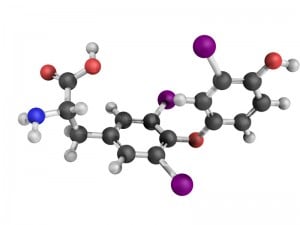
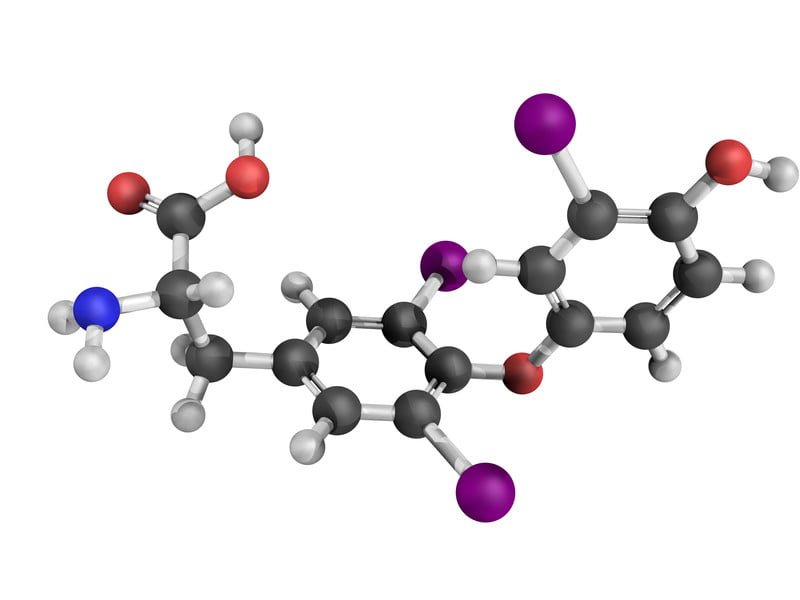
Chemical structure of a T3 molecule, thyroid hormone that affects growth and development, metabolism, body temperature, and heart rate. Relationship between hypothyroidism and celiac disease. Autoimmune thyroiditis is an associated disorder of celiac disease. Thyroid status should be assessed at diagnosis of celiac disease, In the reverse, at diagnosis of thyroid disease, celiac disease should be looked for because untreated hypothyroidism causing constipation and weight gain may hide weight loss and/or diarrhea due to celiac disease that may become evident only after treatment with thyroid hormone.3
- Relationship between hypothyroidism and diagnosis of celiac disease. The rapid identification of celiac disease in thyroid disease is clinically important not only for the high risk of complications inherent to untreated celiac disease, but also because celiac disease is a cause of substitute hormonal therapy failure (thyroxine) in patients with autoimmune thyroid disease.4
- Relationship between hypothyroidism and impaired treatment. The need to use high LT4 (thyroxine medication) doses in the substitutional treatment of hypothyroidism is often the very first sign of one of the pathologies that are connected with malabsorption syndrome, which might have been asymptomatic and undiagnosed previously. Patients who require more than 2 μg/kg body weight of LT4 per day, with constantly increased thyrotropin level, should be diagnosed with the suspicion of an absorption disorder. In celiac disease, a gluten-free diet usually allows a normalization of the need for LT4.5
- Relationship between hypothyroidism and risk factors. A study investigating the natural course of pediatric patients with Hashimoto’s thyroiditis and hyperthyrotropinaemia and looking for possible prognostic factors found that celiac disease, elevated thyroid stimulating hormone (TSH) and antithyroid antibodies at presentation and a progressive increase in thyroid stimulating hormone are predictive factors for thyroid failure in Hashimoto’s thyroiditis patients.6
- Relationship between hypothyroidism and selenium. Selenium deficiency caused by malabsorption in celiac disease may be important in this association. In celiac disease, and autoimmune thyroid disease as well, there is an over production of inflammatory interleukin-15 (IL-15) which inhibits the correct removal of damaged intraepithelial lymphocytes caused by the reaction to gluten. On the reverse, selenium proteins promote the removal of damaged cells. Therefore, selenium deficiency is a key factor directly leading to thyroid and intestinal damage because of the failure to remove cells affected by oxidative damage. Serum levels of IL-15 are directly correlated with the seriousness of tissue damage.7
- Relationship between hypothyroidism and autoimmunity. The association between celiac disease and other immune disorders may be due to the sharing of a common genetic background, such as HLA antigens. However, in a very large study, involving 909 patients with celiac disease, Ventura and his associates found that the development of immune disorders in celiac disease was clearly related to the duration of exposure to gluten.8
How Prevalent Is Autoimmune Thyroiditis In Celiac Disease and/or Gluten Sensitivity?
- A cross-sectional prevalence study in Brazil investigating celiac disease occurrence in autoimmune thyroiditis patients found that the prevalence of celiac disease among patients with autoimmune thyroid disease was 9.3%.9
- A 10 year study found auto-immune thyroid disease in 4.3% of patients with dermatitis herpetiformis and 6.0% with celiac disease.10
- In 136 consecutive patients in Turkey with newly diagnosed autoimmune thyroiditis 5.9% of patients were positive for IgA tissue transglutaminase antibody.11
- In patients who were diagnosed with celiac disease at a hospital endocrinology department in India, 28% had hypothyroidism and 17% had subclinical hypothyroidism.12
What Are The Symptoms Of Autoimmune Thyroiditis?
Autoimmune thyroiditis causing hypothyroidism is marked by these symptoms according to frequency:
- Lowered basal metabolism with weight gain in most people affected.
- Weakness.
- Dry skin and hair with loss of outer third of eyebrows.
- Coarse skin.
- Fatigue or lethargy that interferes with normal activity.
- Mental apathy that causes loss of interest in normal activity.
- Sluggishness and slowed speech.
- Intolerance of cold.
- Decreased sweating.
- Cold skin.
- Thickened tongue.
- Edema of the face causing coarsening of features due to non-pitting edema. Face and head appears larger than normally.
- Pale skin. Pale lips.
- Yellowing of skin, called carotenemia, more noticeable on palms and soles caused by accumulation of carotene in the epidermal layers due to a defect in the conversion of carotene to vitamin A in the liver.
- Memory impairment that progesses.
- Impaired comprehension and disintest in learning.
- Constipation.
- Poor appetite.
- Muscle aches and stiffness.
- Joint pains.
- Clumsiness.
- Hoarseness.
- Dysphagia (difficulty swallowing) occasionally due to pressure from goiter.
- Dry, thin nails. Possibly yellow nails.
- Sadness or depression but pleasant behavior.
- Shortness of breath on exertion.
- Peripheral neuropathy (pain, numbness) due to edema.
- Possible carpal-tunnel syndrome at the wrist due to swelling and compression of the median nerve.
- Possible Restless Leg Syndrome.
- Possible deafness of the perceptive type.
- In females, heavier than normal and irregular menstrual periods and miscarriage.
- Increased risk of infertility in both sexes.
A noted above, myxedema may develop with progression of hypothyroidism especially in the presence of anemia, infection or trauma. Myxedema coma is a life-threatening complication when thyroid hormone gets too low and must be treated in a hospital. Symptoms and signs of myxedema coma include:
- Hypothermia (below normal body temperature).
- Disorientation progressing to unresponsiveness.
- Uncharacteristic moods.
- All over body swelling with tongue thickening and puffy eyes.
- Difficulty breathing with decreased respirations such as 12 breaths per minute.
- Slow pulse such as 50 beats per minute, conduction abnormalities and low blood pressure progressing to heart failure.
- Low blood sugar (hypoglycemia).
How Does Autoimmune Thyroiditis Develop In Celiac Disease and/or Gluten Sensitivity?
- Autoimmune thyroiditis results from production of anti-thyroid antibodies, sharing a common immunopathogenesis with celiac disease. In both celiac disease and autoimmune thyroid disease the genetic markers known as HLA-DQ2 and HLA-DQ8 are overexpressed.
- In celiac disease, and autoimmune thyroid disease as well, there is an over production of inflammatory interleukin-15 (IL-15) which inhibits the correct removal of damaged intraepithelial lymphocytes caused by the reaction to gluten. On the reverse, selenium proteins promote the removal of damaged cells. Therefore, selenium deficiency is a key factor directly leading to thyroid and intestinal damage because of the failure to remove cells affected by oxidative damage. Serum levels of IL-15 are directly correlated with the seriousness of tissue damage.7
- The rate of thyrotoxicosis is significantly elevated in patients with vitamin D deficiency.13
Does Autoimmune Thyroiditis Respond To Gluten-Free Diet?
Response of celiac disease-related autoimmune thyroiditis to gluten free diet varies.14 However, early diagnosis and institution of a gluten free diet may prevent the development of autoimmune thyroid disease.7
CAUTION: Iodine is an essential mineral for the thyroid. However, people with Hashimoto’s disease may be sensitive to harmful side effects from iodine. Taking iodine drops or eating foods containing large amounts of iodine—such as seaweed, dulse, or kelp—may cause or worsen hypothyroidism.2
6 Steps To Improve Autoimmune Thyroiditis In Celiac Disease and/or Gluten Sensitivity:
- [dropcap]1[/dropcap]Remove the Trigger. Maintain a Strict, Nutritious Gluten Free Diet:
[box type=”shadow” ]Treatment. This condition responds to the complete elimination of gluten, which is the required treatment that improves both autoimmune thyroiditis and gut health.
- Gut health is the foundation to restore ALL health. Restored health will enable you to maintain a strict gluten free diet, just as other life tasks will be easier.
- A strict gluten free diet means removing 100% of wheat, barley, rye and oats from the diet.
- Cutting out bread and other obvious sources of gluten is not good enough for recovery. Even 1/8th teaspoon of flour or bread crumb is enough to sustain the inflammation that is damaging your small intestine, causing increased permeability (leaky gut) and allowing undigested gluten to enter your body where it can damage structures and function, and instigate immune inflammatory responses.
Correct Your Individual Nutritional Needs.
- Eat foods that can replenish missing nutrients. Find them under NUTRIENT DEFICIENCIES.
- Take nutritional supplements as needed. Find them under NUTRIENT DEFICIENCIES.
Recovery. You should begin to feel better within a week and notice more energy as inflammation subsides and the absorbing cells that make up the surface lining of your small intestine are better able to function.
- Intestinal lining cells are replaced every 5 days. The healing process is like sunburn where the damaged surface layer of skin sloughs off and is replaced with new normal cells.
- Leaky gut normally resolves in two month after starting a gluten free diet and brings about a big improvement in health. Improvement in intestinal permeability precedes morphometric recovery (cell appearance and structure) of the small intestine in celiac disease.15
- The intestinal lining may take up to a year to heal.[/box]
- [dropcap]2[/dropcap] Reduce Inflammation. Foods to Eat and Foods Not to Eat:
Because gluten is inflammatory, eliminate OTHER inflammatory foods from your diet to reduce an additive effect to gluten. At the same time, try to eat foods that reduce inflammation (anti-inflammatory).
[box type=”shadow” ]Here Are Major Inflammatory Food Types That Reduce Healing:
- Damaging Foods. In susceptible persons, includes corn, dairy (cow), and soy. Lactose, the sugar in any animal milk disrupts intestinal permeability causing leaky gut.16
- Allergenic Foods. Includes foods that trigger the immune sytem to produce IgE antibodies. Allergy testing is the usual way to discover these offending foods.
- Shelf Stable Processed Foods. Includes any that contain additives and preservatives. Look for them on the nutrition label of the box or package. Additives and preservatives also disrupt intestinal permeability causing leaky gut.16
- Fats. Limit deep fried foods, trans-fats, saturated fats (animal fat/butter), and EXCESSIVE omega-6 fatty acid oils like corn oil. Rancid fats, sodium caprate (a medium chain fat), and sucrose monester fatty acid (a food grade surfactant) induce significant disruption of the intestinal barrier that causes leaky gut.16.
- Excessive Refined White Flours (bran layer removed). Includes products made from them such as cookies, bread, cakes, pies. Bran contains the vitamins and minerals that metabolize grains and slows the otherwise rapid entry of sugar from their digestion into the bloodstream. Also disrupt intestinal permeability causing leaky gut.16
- Refined Sugars. Includes white sugar, corn fructose and high fructose corn syrup.
- Certain Spices. Includes paprika and cayenne pepper which disrupt intestinal permeability causing leaky gut.16
- Alcohol and Caffeine. Disrupt intestinal permeability causing leaky gut.16[/box]
[box type=”shadow” ]Here Are Important Anti-Inflammatory Food Types to Promote Health:
- Fruits. Contain ample amounts of vitamins, minerals and phytochemicals which are naturally occuring components in plants that detoxify toxins, carcinogens (reducing the risk by 50%) and mutagens.
- Non-Starchy Vegetables. Support intestinal integrity and provide ample amounts of vitamins, minerals and phytochemicals. Includes green leafy vegetables such as lettuce and kale, also onion, broccoli, garlic, and others.
- High Quality Complex Carbohydrates. Provide vitamins, minerals, and fiber while boosting serotonin levels to help you relax and feel calm. Includes whole grains, legumes, and root vegetables such as carrots, parsnips, sweet potatoes, turnips, red beets, and others.
- Antioxidants. Protect the body from inflammatory oxidant molecules that continually occur and help us handle stress and reduce irritability. Includes vitamin C-containing foods such as lemon, grapefruit, apricot, Brussels sprouts and strawberries, and others. Also, includes vitamin E-containing foods such as nuts, seeds, avocado, olive oil, and others. Cocoa is good, too.
- Omega-3 Fatty Acids. Balance opposing omega-6 fatty acids and bad fats. Fish sources includes tuna, salmon, cod, and others. Plants sources include flax, chia seeds, canola oil, and others.
- Probiotics. Supply normal microbes needed for colon health and health of the body such as these fermented foods: yogurt, kefir, and unpasteurized apple cider vinegar.
- Prebiotics/ High Fiber Foods. Food with fiber keeps our population of colonic microbes healthy.
- Protective Herbs and Spices. See below #6 below for examples.[/box]
- [dropcap]3[/dropcap] Information Sheet You Can Take to Your Doctor or Other Health Professional:
Click here.
- [dropcap]4[/dropcap] Manage Your Medications Safely:
[box type=”shadow” ]
Certain medications used to treat hypothyroidism resulting from autoimmune thyroiditis deplete calcium. Ask your doctor or pharmacist about this possible adverse effect if you are taking any of the drugs listed below. Do not stop prescribed medications without supervision.
THYROID MEDICATION
- Synthroid® depletes calcium.
Many medications deplete selenium and/or vitamin D which are required for normal thyroid function. Here are some:
ANTACIDS / ULCER MEDICATIONS
- Pepcid®, Tagamet®, Zantac® deplete Vitamin D.
- Magnesium and Aluminum Antacid preparations (Gaviscon®, Maalox®, Mylanta®) deplete Vitamin D.
ANTI-INFLAMMATORIES disrupt intestinal permeability which complicates celiac disease.
- Corticosteroids (Prednisone, Medrol®, Aristocort®, Decadron) deplete Vitamin D, Selenium.
ANTICONVULSANTS
- Phenobarbital and Barbituates; and Dilantin®, Tegretol®, Mysoline®, Depakane/Depacon® deplete Vitamin D, Selenium.
BRONCHODILATORS
- Inhaled corticosteroid inhalers (Flovent, Pulmicort and others) that are breathed in on a daily basis as a long term therapy to reduce inflammation in airways deplete Vitamin D.
CHOLESTEROL DRUGS
- Colestid® and Questran® deplete Vitamin D.
FEMALE HORMONES disrupt intestinal permeability which complicate celiac disease.
- Oral Contraceptives (Norinyl®, Ortho-Novum®, Triphasil®, and others) deplete Selenium.
Correlation analysis shows significant association between some trace elements and the duration of contraception and body mass index of the participants.17
LAXATIVES
- Metamucil, FiberCon, Citrucel, Colace, Glycolax, Milk of magnesia, Dulcolax deplete: Vitamin D.
WEIGHT LOSS DRUGS THAT BIND FAT also interfere with absorption of some nutrients.
- Zenicol (Orlistat®) depletes Vitamin D.
[/box]
- [dropcap]5[/dropcap]Nutritional Supplements To Help Correct Deficiencies:
[box type=”shadow” ]
The type and quantity of nutritional supplements that may be needed depend on which nutrients are deficient.
- Multivitamin/mineral combination that provides 100% once a day is useful to improve overall nutrient levels. This is a safe dose, but always check with your doctor to avoid interactions with medications.
- Calcium citrate to replace depleted calcium is the best absorbed of calcium supplements. Calcium carbonate is a poor choice.
- Selenium as indicated by blood test.
- Vitamin D3 as indicated by blood test.
Storage Note: Store container tightly sealed, away from heat, moisture and direct light to avoid loss of potency. That is, in a safe kitchen cabinet – not in the bathroom or on the kitchen table.[/box]
- [dropcap]6[/dropcap]Manage Natural Remedies:
[box type=”shadow” ]Hydration:
- Eight glasses of water are recommended per day unless there is a contraindication such as kidney or heart disease. The Institute of Medicine recommends approximately 2.7 liters (91 ounces) of total water, from all beverages and foods, each day for women and 3.7 liters (125 ounces) daily of total water for men.
- If you are thirsty, drink water. Add fresh, squeezed lemon to water. Lemon is anti-inflammatory, alkalizing and provides vitamin C.
- Hydration Test: Urine should be pale yellow. Fingertips should be plump, without pruning but this may not be reliable when fingers are swollen with edema. Lips should be plump, without puckering. The feeling of thirst can be unreliable.
- What is wrong with soda, coffee, tea, and alcohol? These drinks are dehydrating, increase acid, and deplete nutrients.[/box]
[box type=”shadow” ]Carminatives. The following anti-inflammatory plant sources called carminitives help heal the digestive tract. They also tone the digestive muscles which improves peristalsis, thus aiding in the expulsion of gas from the stomach and intestine to relieve digestive colic and gastric discomfort.
Carminative Food Remedies:
- Raspberry.
- Carrot is also a cleansing digestive tonic.
- Grape is also bile stimulating and a cleansing remedy for sluggish digestion and laxative.
- Redbeets also stimulate and improve digestion and are easily digested.
- Cabbage also stimulates and improves digestion and is also a liver decongestant.
- Lettuce also stimulates and improves digestion and is also an alterative, meaning it improves the function of organs involved with the digestion and excretion of waste products to bring about a gradual change.
- Potatoes are antispasmodic (due to atropine like properties) and a liver remedy.
Carminative Herb Remedies:
- Sage is also a digestive, astringent, bile stimulant and energy tonic that heals the mucosa. Drink as tea or use in cooking.
- Chamomile, lemon balm, and fennel, (as a tea) also help relieve nervous tension.
- Parsley also relieves indigestion.
- Rosemary as a tea and in cooking also is a nervous system tonic for stress and fatigue, bile stimulant, and can relieve headaches and indigestion.
- Thyme is also soothing remedy useful for stimulating digestion of rich, fatty foods.
Carminative Spice Remedies:
- Cloves are also antispasmodic.
- Nutmeg is also useful for indigestion.
- Ginger.[/box]
[box type=”shadow” ]Exercise Helps:
Exercise improves circulation and rids the body of toxins.
- Walking is aerobic exercise that reconditions the whole body to improve stamina. Read more about Exercise and Fitness.
- Weight training builds muscle. Read more about Exercise and Fitness.
- Stretching improves flexibilty. Read more about Exercise and Fitness.
Note: Exercise is important, but the amount and type of exercise undertaken depends on your health. Your first priority is to heal. [/box]
What Do Medical Research Studies Tell About Autoimmune Thyroiditis In Celiac Disease and/or Gluten Sensitivity?
RESEARCH STUDY SUMMARIES
“Prevalence and clinical features of celiac disease in patients with autoimmune thyroiditis: cross-sectional study.” This cross-sectional prevalence study investigating celiac disease occurrence in autoimmune thyroiditis patients found that the prevalence of celiac disease among patients with autoimmune thyroid disease was 9.3%. Patients were tested for anti-endomysial and anti-transglutaminase antibodies between August 2010 and July 2011.
Only one patient complained of diarrhea and none presented anemia. The researchers state, “Among at-risk populations, like autoimmune thyroiditis patients, the presence of diarrhea or anemia should not be used as a criterion for indicating celiac disease investigation. This must be done for all autoimmune thyroiditis patients because of its high prevalence.”
Fifty-three patients with autoimmune thyroiditis were included; 92.5% were women, with mean age of 49.0 ± 13.5 years. Five patients (9.3%) were serologically positive for celiac disease: three of them (5.6%) were reactive for anti-endomysial antibodies and two (3.7%) for anti-transglutaminase. None of them exhibited anemia and one presented diarrhea. Endoscopy was performed on two patients: one with normal histology and the other with lymphocytic infiltrate and villous atrophy.18
“Hospital admissions for vitamin D related conditions and subsequent immune-mediated disease: record-linkage studies.” This study investigating the reported association between vitamin D deficiency and the risk of developing immune-mediated diseases showed that patients with vitamin D deficiency may have an increased risk of developing some immune-mediated diseases including thyroid disease, although reverse causality or confounding cannot be ruled out.
Researchers analyzed a database of linked statistical records of hospital admissions and death registrations for the whole of England (from 1999 to 2011). Rate ratios for immune-mediated disease were determined, comparing vitamin D deficient cohorts (individuals admitted for vitamin D deficiency or markers of vitamin D deficiency) with comparison cohorts.
After hospital admission for either vitamin D deficiency, osteomalacia or rickets, there were significantly elevated rates of Addison’s disease, ankylosing spondylitis, autoimmune hemolytic anemia, chronic active hepatitis, celiac disease, Crohn’s disease, diabetes mellitus, pemphigoid, pernicious anemia, primary biliary cirrhosis, rheumatoid arthritis, Sjogren’s syndrome, systemic lupus erythematosus, thyrotoxicosis, and significantly reduced risks for asthma and myxoedema.13
“Endocrine manifestations of celiac disease.” This study investigating the prevalence of endocrinopathies in 36 patients who were diagnosed with celiac disease at a hospital endocrinology department found hypothyroidism in 28% and subclinical hypothyroidism in 17% among a variety of manifestations.12
“The natural history of the normal/mild elevated TSH serum levels in children and adolescents with Hashimoto’s thyroiditis and isolated hyperthyrotropinaemia: a 3-year follow-up.” This retrospective cross-sectional study investigating the natural course of patients with Hashimoto’s thyroiditis and hyperthyrotropinaemia and looking for possible prognostic factors found that celiac disease, elevated thyroid stimulating hormone and antithyroid antibodies at presentation and a progressive increase in thyroid stimulating hormone are predictive factors for thyroid failure in Hashimoto’s thyroiditis patients.
Three hundred and twenty-three patients with Hashimoto’s thyroiditis (88 boys and 235 girls) and 59 with hyperthyrotropinaemia (30 boys and 29 girls), mean age 9·9 ± 3·8 years were included in the study. When first examined, 236 of the children with Hashimoto’s thyroiditis had a normal Hashimoto’s thyroiditis (G0) and in 87, it was elevated but less than 100% of the upper limit (G1). All hyperthyrotropinaemia subjects had elevated thyroid stimulating hormone (TSH). Potential risk factors for thyroid failure were evaluated after 3 years and included the presence or familiarity for endocrine/autoimmune diseases, premature birth, signs and symptoms of hypothyroidism, TSH levels, antithyroid antibodies and thyroid volume.
RESULTS: Hashimoto’s thyroiditis (HT): Of those with HT, 170 normal TSH (GO)patients remained stable, 31 moved to G1 and 35 to G2 (hypothyroidism). Thirty-six G1 children moved to G0, 17 remained stable and 34 moved to G2. Of patients with hyperthyrotropinaemia (IH): 23 normalized, 28 remained stable and eight became overtly hypothyroid. In patients with HT, the presence of celiac disease, elevated TSH and thyroid peroxidase antibodies (TPOAb) increased the risk of developing hypothyroidism by 4·0-, 3·4- and 3·5-fold, respectively. The increase in TSH levels during follow-up was strongly predictive of the development of hypothyroidism. In patients with IH, no predictive factor could be identified.6
“Celiac disease in children with Down syndrome: importance of follow-up and serological screening.” This study investigating the incidence of celiac disease in children with Down syndrome, to assess the availability of IgA AGA and EMA for serologic screening, and to highlight the importance of follow-up demonstrated that children with Down syndrome should be carefully examined in their follow-up, and celiac disease should be considered in cases with growth retardation. All study patients with abnormal biopsy were below the 10th percentile for weight and height. Positivity of both IgA AGA and EMA serologic screening tests gave the most reliable results. Hypothyroidism was detected in one of 11 cases where at least one serologic marker was positive.19
“Thyroid-related autoantibodies and celiac disease: a role for a gluten-free diet?” This study evaluating the presence of celiac disease in 100 patients with autoimmune thyroid disease demonstrated that serologic markers became undetectable 6 months after beginning gluten free diet, but thyroid autoantibodies did not positively correlate with gluten free diet.14
“Diseases associated with dermatitis herpetiformis.” This study investigating the occurrence of associated diseases in a cohort of 305 patients with dermatitis herpetiformis (DH) followed for a mean of 10 years compared with results from a cohort of patients with celiac disease demonstrated autoimmune thyroid disease in 4.3% of DH patients and 6.0% celiac disease patients.10
CASE REPORT SUMMARIES
“Multiple Disease Associations in Autoimmune Polyglandular Syndrome Type II. “ This case report describes the course of a 25 year old female with a history of ulcerative colitis, celiac disease and type 1 diabetes who presented with mental status changes. She was diagnosed with Hashimoto’s encephalopathy and treated with high dose steroids and intravenous immunoglobulin. She recovered well from her encephalopathy but her post-hospitalization course was complicated due to the development of Addison’s disease, vitiligo, sero-negative arthritis, and hypothyroidism.
This patient had a dramatic development of eight autoimmune diseases over the course of ten years. She developed Addison’s disease, hypothyroidism, type 1 diabetes, Hashimoto’s encephalopathy, vitiligo, celiac disease, sero-negative arthritis, and ulcerative colitis. This represents a particularly aggressive course of APS II and this combination of autoimmune diseases has not been previously reported. It highlights the potential complexity and severity of the clinical course of APS II.20
“Multiple immune disorders in unrecognized celiac disease: a case report.” This case report describes the course of a 34 year old female patient with unrecognized celiac disease and multiple extra intestinal manifestations, mainly related to a deranged immune function, including thyroiditis and anti-thyreoglobulin antibodies that disappeared or improved after the implementation of a gluten-free diet.
After six months of controlled gluten free diet, the patient’s body weight increased 12 kg; laboratory investigations demonstrated normalization of serum amylase, serum lipase and immunoglobulin levels; antigliadin, anti-2-glicoprotein-1 and were no longer detectable, but antiendomysial antibodies were still present. Endoscopy showed a normal appearance of duodenal mucosa, and duodenal biopsy revealed a partial recovery of duodenal morphology. After 18 months of gluten-free diet, antiendomysial antibodies disappeared; creatinine clearance increased, but proteinuria further worsened (2.9 g/day, Table 1), and albumin levels were still low. After 24 months of gluten-free diet, a new duodenal biopsy showed complete recovery of villous architecture. Renal function further improved and proteinuria markedly decreased (Table 1). Amylase, lipase, and immunoglobulin levels were within the normal range. Anti-2-glicoprotein-1, anti-thyreoglobulin, antigliadin, antiendomysial and anti-TTG antibodies were undetectable. A coagulation study was normal.
“Although both celiac disease and the other manifestations of a deranged immunity might be explained on the basis of a common genetic predisposition to this kind of disorders, some findings suggest that celiac disease itself is responsible for the initiation of the immunological response. Indeed, persistent stimulation by some proinflammatory cytokines, such as interferon and tumor necrosis factor , could induce further processing of autoantigens and their presentation to T lymphocytes by macrophage-type immunocompetent cells. As a matter of fact, the prevalence of immune diseases among patients with celiac disease seems proportional to the time of exposure to gluten, and many immune alterations disappear following the recognition of celiac disease and appropriate treatment, just as it occurred in this patient.”21
“Unusual association of thyroiditis, Addison’s disease, ovarian failure and celiac disease in a young woman.” This case report describes the course of a 23 year old woman with a diagnosis of hypothyroidism due to Hashimoto’s thyroiditis, autoimmune Addison’s disease, and kariotypically normal spontaneous premature ovarian failure. Search for celiac disease revealed positive EMA antibodies and total villous atrophy at jejunal biopsy.
Marked clinical improvement and a progressive decrease in the need for thyroid and adrenal replacement therapies occurred over a 3-month period. After 6 months serum EMA became negative and after 12 months a new jejunal biopsy showed complete mucosal recovery. After 18 months the anti-thyroid antibodies titre decreased significantly and thyroid substitution therapy was discontinued. The precocious identification of celiac disease in polyglandular disease is clinically relevant not only for the high risk of complications inherent to untreated celiac disease, but also because celiac disease is a cause of substitute hormonal therapy failure in patients with autoimmune thyroid disease.22
Sources:- Taber’s Cyclopedic Medical Dictionary. 19th ed. F.A. Davis Company. Philadelphia, PA. [↩]
- National Endocrine and Metabolic Diseases Information Service. [↩] [↩]
- Stazi AV, Trinti B. Selenium status and over-expression of interleukin-15 in celiac disease and autoimmune thyroid diseases. Ann Ist Super Sanita. 2010;46(4):389-99.DOI: 10.4415/ANN_10_04_06. [↩]
- Valentino R, Savastano S, Tommaselli AP. Unusual association of thyroiditis, Addison’s disease, ovarian failure and celiac disease in a young woman. Journal of Endocrinological Investigation. May 1999;22(5):390-4. [↩]
- Ruchała M, Szczepanek-Parulska E, Zybek A. The influence of lactose intolerance and other gastro-intestinal tract disorders on L-thyroxine absorption. Endokrynol Pol. 2012;63(4):318-23. [↩]
- Radetti G, Maselli M, Buzi F, Corrias A, Mussa A, Cambiaso P, Salerno M, Cappa M, Baiocchi M, Gastaldi R, Minerba L, Loche S. The natural history of the normal/mild elevated TSH serum levels in children and adolescents with Hashimoto’s thyroiditis and isolated hyperthyrotropinaemia: a 3-year follow-up. Clin Endocrinol (Oxf). 2012 Mar;76(3):394-8. doi: 10.1111/j.1365-2265.2011.04251.x. [↩] [↩]
- Stazi AV, Trinti B. Selenium status and over-expression of interleukin-15 in celiac disease and autoimmune thyroid diseases. Ann Ist Super Sanita. 2010;46(4):389-99.DOI: 10.4415/ANN_10_04_06. [↩] [↩] [↩]
- La Villa G, Pantaleo P, Tarquini R, Cirami L, Perfetto F, Mancuso F, Laffi G. Multiple immune disorders in unrecognized celiac disease: a case report. World J Gastroenterol. 2003;9(6):1377-1380. [↩]
- Ventura A, Ronsoni MF, Shiozawa MB, Dantas-Corrêa EB, Canalli MH, Schiavon Lde L, Narciso-Schiavon JL. Prevalence and clinical features of celiac disease in patients with autoimmune thyroiditis: cross-sectional study. Sao Paulo Med J. 2014 Dec;132(6):364-71. [↩]
- Reunala T, Collin P. Diseases associated with dermatitis herpetiformis. British Journal of Dermatology. Mar 1997;136(3):315-8. [↩] [↩]
- Guliter S, Yakaryilmaz F, Ozkurt Z, Ersoy R, Ucardag D, Caglayan O, Atasoy P. Prevalence of coeliac disease in patients with autoimmune thyroiditis in a Turkish population. World J Gastroenterol. 2007 Mar 14;13(10):1599-601. [↩]
- Philip R, Patidar P, Saran S, Agarwal P, Gupta K. Endocrine manifestations of celiac disease. Indian J Endocrinol Metab. 2012 December; 16(Suppl 2): S506–S508. [↩] [↩]
- Ramagopalan SV, Goldacre R, Disanto G, Giovannoni G, Goldacre MJ. Hospital admissions for vitamin D related conditions and subsequent immune-mediated disease: record-linkage studies. BMC Med. 2013 Jul 25;11:171. doi: 10.1186/1741-7015-11-171. [↩] [↩]
- Mainardi E, Montanelli A, Dotti M, Nano R, Moscato G. Thyroid-related autoantibodies and celiac disease: a role for a gluten-free diet? Journal of Clinical Gastroenterology. Sep 2002;35(3):245-8. [↩] [↩]
- Cummins AG, Thompson FM, Butler RN, et al. Improvement in intestinal permeability precedes morphometric recovery of the small intestine in coeliac disease. Clinical Science. Apr 2001;100(4):379-86. [↩]
- Farhadi A, Banan A, Fields J, Keshavarzian A. Intestinal barrier: an interface between health and disease. Journal of Gastroenterology and Hepatology. 2003;18:479-91. [↩] [↩] [↩] [↩] [↩] [↩]
- Akinloye O1, Adebayo TO, Oguntibeju OO, Oparinde DP, Ogunyemi EO. Effects of contraceptives on serum trace elements, calcium and phosphorus levels. West Indian Med J. 2011 Jun;60(3):308-15. [↩]
- Ventura A, Ronsoni MF, Shiozawa MB, Dantas-Corrêa EB, Canalli MH, Schiavon Lde L, Narciso-Schiavon JL. Prevalence and clinical features of celiac disease in patients with autoimmune thyroiditis: cross-sectional study. Sao Paulo Med J. 2014 Dec;132(6):364-71. Epub 2014 Sep 2. [↩]
- Cogulu O, Ozkinay F, Gunduz C, et al. Celiac disease in children with Down syndrome: importance of follow-up and serological screening. Pediatrics International: Official Journal of the Japan Pediatric Society. Aug 2003;45(4):395-9. [↩]
- Maturu A, Michels A, Draznin B. Multiple Disease Associations in Autoimmune Polyglandular Syndrome Type II. with autoimmune polyglandular syndrome type II (APS II). Endocr Pract. 2014 Aug 22:1-13. [↩]
- La Villa G, Pantaleo P, Tarquini R, Cirami L, Perfetto F, Mancuso F, Laffi G. Multiple immune disorders in unrecognized celiac disease: a case report. World J Gastroenterol 2003; 9(6): 1377-1380. [↩]
- Valentino R, Savastano S, Tommaselli AP. Unusual association of thyroiditis, Addison’s disease, ovarian failure and celiac disease in a young woman. Journal of Endocrinological Investigation. May 1999;22(5):390-4. [↩]