Page Contents
What Is Dermatomyositis?
Dermatomyositis is a rare autoimmune systemic disease of the connective tissue that is characterized by inflammatory and debilitating degenerative changes in the muscles and in the skin.
Dermatomyositis results in symmetric, proximal muscle weakness of limbs (upper arms and legs), and skin manifestations. 50-70% of patients have circulating myositis-specific auto-antibodies.
The course of dermatomyositis is unpredictable being marked by spontaneous flare-ups and remissions. It can begin slowly or abruptly according to the factor that is triggering the onset such as infection, medications like phenytoin, and autoimmune disease.
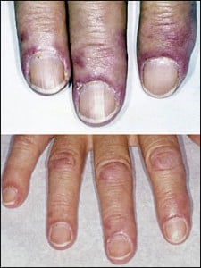
Q: What are the skin manifestations of dermatomyositis?
A: Classic skin manifestations of dermatomyositis include these features:
- The heliotrope rash (lilac color) on upper eyelids.
- Rash on face, neck, shoulders, upper chest, elbows, knees, knuckles, and back.
- Gottron’s papules (scaly, red eruptions or patches over the knuckles, elbows, and knees).
- The V-sign (rash front of neck and chest).
- The shawl sign (rash distribution on shoulders and back).1
Additional cutaneous manifestations are described below under symptoms.
Dermatomyositis is associated with an increased risk of cancer, other autoimmune diseases, such as lupus and psoriasis, and it can be a complication of interferon-α therapy. About 1 person in 100,000 are affected according to various studies. While it affects all ages, women have twice the occurence of men.
There is no cure for dermatomyositis, but the symptoms can be treated. Options include medication, physical therapy, exercise, heat therapy (including microwave and ultrasound), orthotics and assistive devices, and rest. The standard treatment for dermatomyositis is a corticosteroid drug, given either in pill form or intravenously. Immunosuppressant drugs, such as azathioprine and methotrexate, may reduce inflammation in people who do not respond well to prednisone.2
What Is Dermatomyositis In Celiac Disease and/or Gluten Sensitivity?
- Relationship between dermatomyositis and celiac disease. Dermatomyositis is an auotimmune disorder associated with celiac disease.
- Relationship between dermatomyositis and gluten. The association between deliac disease and other immune disorders may be due to the sharing of a common genetic background, such as HLA antigens. However, in a very large study, involving 909 patients with celiac disease, Ventura and his associates found that the development of immune disorders in celiac disease was clearly related to the duration of exposure to gluten.3
How Prevalent Is Dermatomyositis In Celiac Disease and/or Gluten Sensitivity?
The association between dermatomyositis and celiac disease in children has been well documented. In the adult population, however, the association has not been clearly established,4 except by case reports documenting the co-existence in individual patients.5
A study investigating the presence of celiac disease and antibodies associated with celiac disease in 51 adult patients with inflammatory myopathies and their relationship found that celiac disease is more prevalent in patients with inflammatory myopathies than in the general population.6
What Are The Symptoms Of Dermatomyositis?
Dermatomyositis is marked by these symptoms:
- Gottron rash. Reddish patches, sometimes raised, smooth or scaly, that forms defined areas over bony prominences like the elbow.
- Heliotope rash. A purplish lilac discoloration that affects the eyelids.
- Poikiloderma. A chronic reddish-brown rash on the cheeks and neck.
- An itchy rash. A flat red eruption on the face. forehead, and chest that can be triggered by sunlight and may cover much of the body.
- Nail involvement called periungual telangiectasias (rash around nails (see photo upper left).
- Nail cuticle overgrowth.
- “Mechanic’s hands” (dry, cracked, rough looking).
- Wasting of skeletal muscles, especially of the shoulder and pelvic girdles causing weakness with difficulty climbing stairs, rising from sqatting position, and reaching overhead.
- Muscle ache and tenderness.
- Difficulty breathing from lung involvement.
- Alopecia, thin hair.
- Calcinosis (calcium deposits in skin). Calcium deposits appear as hard bumps under the skin or in the muscle. Calcinosis most often occurs 1-3 years after the disease begins and is associated with damaged, inflamed, or necrotic skin.
How Does Dermatomyositis Develop In Celiac Disease and/or Gluten Sensitivity?
- Dermatomyositis results from an immune mechanism not well understood.
Does Dermatomyositis Respond To Gluten-Free Diet?
Yes. After a patient was put on a strict gluten-free diet, both nutritional deficiencies and the dermatomyositis resolved.7
6 Steps To Improve Dermatomyositis In Celiac Disease and/or Gluten Sensitivity:
- 1Remove the Trigger. Maintain a Strict, Nutritious Gluten Free Diet:
- Gut health is the foundation to restore ALL health. Restored health will enable you to maintain a strict gluten free diet, just as other life tasks will be easier.
- A strict gluten free diet means removing 100% of wheat, barley, rye and oats from the diet.
- Cutting out bread and other obvious sources of gluten is not good enough for recovery. Even 1/8th teaspoon of flour or bread crumb is enough to sustain the inflammation that is damaging your small intestine, causing increased permeability (leaky gut) and allowing undigested gluten to enter your body where it can damage structures and function, and instigate immune inflammatory responses.
Correct Your Individual Nutritional Needs.
- Eat foods that can replenish missing nutrients. Find them under NUTRIENT DEFICIENCIES.
- Take nutritional supplements as needed. Find them under NUTRIENT DEFICIENCIES.
Recovery. You should begin to feel better within a week and notice more energy as inflammation subsides and the absorbing cells that make up the surface lining of your small intestine are better able to function.
- Intestinal lining cells are replaced every 5 days. The healing process is like sunburn where the damaged surface layer of skin sloughs off and is replaced with new normal cells.
- Leaky gut normally resolves in two month after starting a gluten free diet and brings about a big improvement in health. Improvement in intestinal permeability precedes morphometric recovery (cell appearance and structure) of the small intestine in celiac disease.8
- The intestinal lining may take up to a year to heal.
- 2 Reduce Inflammation. Foods to Eat and Foods Not to Eat:
Because gluten is inflammatory, eliminate OTHER inflammatory foods from your diet to reduce an additive effect to gluten. At the same time, try to eat foods that reduce inflammation (anti-inflammatory).
- Damaging Foods. In susceptible persons, includes corn, dairy (cow), and soy. Lactose, the sugar in any animal milk disrupts intestinal permeability causing leaky gut.9
- Allergenic Foods. Includes foods that trigger the immune sytem to produce IgE antibodies. Allergy testing is the usual way to discover these offending foods.
- Shelf Stable Processed Foods. Includes any that contain additives and preservatives. Look for them on the nutrition label of the box or package. Additives and preservatives also disrupt intestinal permeability causing leaky gut.9
- Fats. Limit deep fried foods, trans-fats, saturated fats (animal fat/butter), and EXCESSIVE omega-6 fatty acid oils like corn oil. Rancid fats, sodium caprate (a medium chain fat), and sucrose monester fatty acid (a food grade surfactant) induce significant disruption of the intestinal barrier that causes leaky gut.9.
- Excessive Refined White Flours (bran layer removed). Includes products made from them such as cookies, bread, cakes, pies. Bran contains the vitamins and minerals that metabolize grains and slows the otherwise rapid entry of sugar from their digestion into the bloodstream. Also disrupt intestinal permeability causing leaky gut.9
- Refined Sugars. Includes white sugar, corn fructose and high fructose corn syrup.
- Certain Spices. Includes paprika and cayenne pepper which disrupt intestinal permeability causing leaky gut.9
- Alcohol and Caffeine. Disrupt intestinal permeability causing leaky gut.9
- Fruits. Contain ample amounts of vitamins, minerals and phytochemicals which are naturally occuring components in plants that detoxify toxins, carcinogens (reducing the risk by 50%) and mutagens.
- Non-Starchy Vegetables. Support intestinal integrity and provide ample amounts of vitamins, minerals and phytochemicals. Includes green leafy vegetables such as lettuce and kale, also onion, broccoli, garlic, and others.
- High Quality Complex Carbohydrates. Provide vitamins, minerals, and fiber while boosting serotonin levels to help you relax and feel calm. Includes whole grains, legumes, and root vegetables such as carrots, parsnips, sweet potatoes, turnips, red beets, and others.
- Antioxidants. Protect the body from inflammatory oxidant molecules that continually occur and help us handle stress and reduce irritability. Includes vitamin C-containing foods such as lemon, grapefruit, apricot, Brussels sprouts and strawberries, and others. Also, includes vitamin E-containing foods such as nuts, seeds, avocado, olive oil, and others. Cocoa is good, too.
- Omega-3 Fatty Acids. Balance opposing omega-6 fatty acids and bad fats. Fish sources includes tuna, salmon, cod, and others. Plants sources include flax, chia seeds, canola oil, and others.
- Probiotics. Supply normal microbes needed for colon health and health of the body such as these fermented foods: yogurt, kefir, and unpasteurized apple cider vinegar.
- Prebiotics/ High Fiber Foods. Food with fiber keeps our population of colonic microbes healthy.
- Protective Herbs and Spices. See below #6 below for examples.
- 3 Information Sheet You Can Take to Your Doctor or Other Health Professional:
- 4 Manage Your Medications Safely:
Certain medications used to treat dermatomyositis cause nutritional deficiencies that can promote complications in celiac disease. Ask your doctor or pharmacist about this possible adverse effect if you are taking any of the drugs listed below. Do not stop prescribed medications without supervision.
This is not a complete listing.
ANTI-INFLAMMATORIES disrupt intestinal permeability which complicates celiac disease.
- Corticosteroids for inflammation (Prednisone, Medrol®, Aristocort®, Decadron) deplete Calcium, Vitamin D, Magnesium, Zinc, Vitamin C, Vitamin B6, Vitamin B12, Folic Acid, Selenium, Chromium, Phosphorus.
- NSAIDS for pain (Motrin®, Aleve®, Advil®, Anaprox®, Dolobid®, Feldene®, Naprosyn® and others) deplete Folic acid.
- 5Nutritional Supplements To Help Correct Deficiencies:
The type and quantity of nutritional supplements that may be needed depend on which nutrients are deficient.
- Multivitamin/mineral combination that provides 100% once a day is useful to improve overall nutrient levels. This is a safe dose, but always check with your doctor to avoid interactions with medications.
- B-Complex with 100% to 300% or as as prescribed.
- Calcium citrate is the best absorbed of calcium supplements. Calcium carbonate is a poor choice.
- Vitamin D3 as prescribed following blood test for status.
- Chelated magnesium as prescribed but do not take at same time as calcium because they compete for absorption.
- Zinc as prescribed following blood test for status.
- Selenium as prescribed following blood test for status.
- Chromium as prescribed following blood test for status.
Storage Note: Store container tightly sealed, away from heat, moisture and direct light to avoid loss of potency. That is, in a safe kitchen cabinet – not in the bathroom or on the kitchen table.
- 6Manage Natural Remedies:
- Eight glasses of water are recommended per day unless there is a contraindication such as kidney or heart disease. The Institute of Medicine recommends approximately 2.7 liters (91 ounces) of total water, from all beverages and foods, each day for women and 3.7 liters (125 ounces) daily of total water for men.
- If you are thirsty, drink water. Add fresh, squeezed lemon to water. Lemon is anti-inflammatory, alkalizing and provides vitamin C.
- Hydration Test: Urine should be pale yellow. Fingertips should be plump, without pruning but this may not be reliable when fingers are swollen with edema. Lips should be plump, without puckering. The feeling of thirst can be unreliable.
- What is wrong with soda, coffee, tea, and alcohol? These drinks are dehydrating, increase acid, and deplete nutrients.
Carminative Food Remedies:
- Raspberry.
- Carrot is also a cleansing digestive tonic.
- Grape is also bile stimulating and a cleansing remedy for sluggish digestion and laxative.
- Redbeets also stimulate and improve digestion and are easily digested.
- Cabbage also stimulates and improves digestion and is also a liver decongestant.
- Lettuce also stimulates and improves digestion and is also an alterative, meaning it improves the function of organs involved with the digestion and excretion of waste products to bring about a gradual change.
- Potatoes are antispasmodic (due to atropine like properties) and a liver remedy.
Carminative Herb Remedies:
- Sage is also a digestive, astringent, bile stimulant and energy tonic that heals the mucosa. Drink as tea or use in cooking.
- Chamomile, lemon balm, and fennel, (as a tea) also help relieve nervous tension.
- Parsley also relieves indigestion.
- Rosemary as a tea and in cooking also is a nervous system tonic for stress and fatigue, bile stimulant, and can relieve headaches and indigestion.
- Thyme is also soothing remedy useful for stimulating digestion of rich, fatty foods.
Carminative Spice Remedies:
- Cloves are also antispasmodic.
- Nutmeg is also useful for indigestion.
- Ginger.
Exercise improves circulation and rids the body of toxins.
- Walking is aerobic exercise that reconditions the whole body to improve stamina. Read more about Exercise and Fitness.
- Weight training builds muscle. Read more about Exercise and Fitness.
- Stretching improves flexibilty. Read more about Exercise and Fitness.
Note: Exercise is important, but the amount and type of exercise undertaken depends on your health. Your first priority is to heal.
What Do Medical Research Studies Tell About Dermatomyositis In Celiac Disease and/or Gluten Sensitivity?
CASE REPORT SUMMARIES
“Calcinosis cutis: A rare feature of adult dermatomyositis.” This case report describes calcinosis in a 55-year-old woman with dermatomyositis who presented with dystrophic calcinosis resistant to medical treatment. Calcinosis cutis is the deposition of insoluble calcium salts in the skin. In connective tissue diseases, calcinosis is mostly of the dystrophic type and it seems to be a localized process rather than an imbalance of calcium homeostasis. Calcium deposits may be intracutaneous, subcutaneous, fascial, or intramuscular.
The woman was referred for evaluation because of multiple, firm nodules of the lateral hips since 1994. At that time, dermatomyositis was diagnosed based on cutaneous, muscular and pulmonary involvement. The nodules, gradually enlarging since 1999, have begun to cause incapacitation pain and many exude a yellowish material suggestive of calcium. She denied an inciting traumatic event. Combinations of oral prednisone, hydroxychloroquine, or chloroquine, have been able to control the heliotrope rash, Gottron papules, and myositis, but have not prevented progression of nodule formation.10
“Celiac disease and antibodies associated with celiac disease in patients with inflammatory myopathy.” This study investigating the presence of celiac disease and antibodies associated with celiac disease in patients with inflammatory myopathies and their relationship found that celiac disease is more prevalent in patients with inflammatory myopathies than in the general population.
Serum antigliadin, anti-tissue transglutaminase, and antiendomysial antibodies were determined in 51 patients with inflammatory myopathies. HLA-DQ2 and -DQ8 alleles were studied to assess their complementary diagnostic value. Jejunal biopsy was performed in patients with moderate to high levels of antigliadin antibodies. Patients with jejunal histology consistent with celiac disease initiated a gluten-free diet. Seventeen patients (31%) were positive for antigliadin antibodies, which were significantly more frequent in patients with inclusion-body myositis than dermatomyositis. Positive status to HLA-DQ2 and/or -DQ8 did not differ between antigliadin-positive (75% and 12.5%) or -negative (60% and 15%) patients. Three of five jejunal biopsies were diagnostic for celiac disease with histological normalization after a gluten-free diet.
The diagnostic value of HLA-DQ2 or -DQ8 haplotypes to detect celiac disease in patients with inflammatory myopathy is limited.6
“Dermatomyositis associated with celiac disease: response to a gluten-free diet.” This case report describes concomitant dermatomyositis and celiac disease in a 40-year-old woman. After having been diagnosed with dermatomyositis and iron deficiency anemia, this patient was referred to the gastroenterology clinic to exclude a gastrointestinal malignancy. Blood tests revealed various vitamin deficiencies consistent with malabsorption. The results of gastroscopy with duodenal biopsy were consistent with celiac disease. After she was put on a strict gluten-free diet, both nutritional deficiencies and the dermatomyositis resolved. The patient’s human leukocyte antigen haplotype study was positive for DR3 and DQ2 genotypes, which have been shown to be associated with both juvenile dermatomyositis and celiac disease. It is suggested that patients with newly diagnosed dermatomyositis be investigated for concomitant celiac disease even in the absence of gastrointestinal symptoms.4
”An uncommon association: celiac disease and dermatomyositis in adults.” This case report of a patient with malabsorption after a 2 year history of dermatomyositis describes subsequent diagnosis of celiac disease. Celiac disease should be suspected in patients with dermatomyositis who exhibit a malabsorption syndrome. Evaluation for celiac disease, including anti-gliadin antibodies, anti-endomysium antibodies, and tissue transglutaminase antibodies should be considered in dermatomyositis patients presenting with unusual and unexplained gastrointestinal features.5
- Marvi U, Chung L, Fiorentino DF. Clinical presentation and evaluation of dermatomyositis. Indian J Dermatol. 2012 Sep;57(5):375-81. doi: 10.4103/0019-5154.100486. [↩]
- National Institute of Neurological Disorders and Stroke. [↩]
- La Villa G, Pantaleo P, Tarquini R, Cirami L, Perfetto F, Mancuso F, Laffi G. Multiple immune disorders in unrecognized celiac disease: a case report. World J Gastroenterol. 2003;9(6):1377-1380. [↩]
- Song MS, Farber D, Bitton A, Jass J, Singer M, Karpati G. Dermatomyositis associated with celiac disease: response to a gluten-free diet. Can J Gastroenterol. 2006 Jun;20(6):433-5. [↩] [↩]
- Marie I, Lecomte F, Hachulla E, et al. An uncommon association: celiac disease and dermatomyositis in adults. Clinical and Experimental Rheumatology. Mar-Apr 2001;19(2):201-3. [↩] [↩]
- Selva-O’Callaghan A, Casellas F, de Torres I, Palou E, Grau-Junyent JM, Vilardell-Tarrés M. Celiac disease and antibodies associated with celiac disease in patients with inflammatory myopathy. Muscle Nerve. 2007 Jan;35(1):49-54. [↩] [↩]
- Song MS, Farber D, Bitton A, Jass J, Singer M, Karpati G. Dermatomyositis associated with celiac disease: response to a gluten-free diet. Can J Gastroenterol. 2006 Jun;20(6):433-5. [↩]
- Cummins AG, Thompson FM, Butler RN, et al. Improvement in intestinal permeability precedes morphometric recovery of the small intestine in coeliac disease. Clinical Science. Apr 2001;100(4):379-86. [↩]
- Farhadi A, Banan A, Fields J, Keshavarzian A. Intestinal barrier: an interface between health and disease. Journal of Gastroenterology and Hepatology. 2003;18:479-91. [↩] [↩] [↩] [↩] [↩] [↩]
- Lobo IM, Machado S, Teixeira M, Selores M. Calcinosis cutis: A rare feature of adult dermatomyositis. Dermatology Online Journal 2008;14 (1): 10. [↩]